Bringing you the latest news in Biology. The natural science concerned with the study of life and living organisms, including their structure, function, growth, evolution, distribution, identification and taxonomy.
According to Medical Xpress (This article and its images were originally posted on Medical Xpress September 26, 2018 at 04:54PM.)
An estimated 80,000 Americans died of flu and its complications last winter—the disease’s highest death toll in at least four decades.
The director of the Centers for Disease Control and Prevention, Dr. Robert Redfield, revealed the total in an interview Tuesday night with The Associated Press.
Flu experts knew it was a very bad season, but at least one found the size of the estimate surprising.
“That’s huge,” said Dr. William Schaffner, a Vanderbilt University vaccine expert. The tally was nearly twice as much as what health officials previously considered a bad year, he said.
In recent years, flu-related deaths have ranged from about 12,000 to 56,000, according to the CDC.
Last fall and winter, the U.S. went through one of the most severe flu seasons in recent memory. It was driven by a kind of flu that tends to put more people in the hospital and cause more deaths, particularly among young children and the elderly.
The season peaked in early February and it was mostly over by the end of March.
Got any news, tips or want to contact us directly? Feel free to email us: esistme@gmail.com.
To see more posts like these; please subscribe to our newsletter. By entering a valid email, you’ll receive top trending reports delivered to your inbox.
__
This article and its images were originally posted on [Medical Xpress] September 26, 2018 at 04:54PM. Credit to the original author and Medical Xpress | ESIST.T>G>S Recommended Articles Of The Day.
Donations are appreciated and go directly to supporting ESIST.Tech. Thank you in advance for helping us to continue to be a part of your online entertainment!
According to Science and technology (This article and its images were originally posted on Science and technology September 20, 2018 at 10:51AM.)
print-edition icon Print edition | Science and technology
CELLS divide many times throughout their lives. But they cannot do it indefinitely. Once they have reached the limits of their reproductive powers, they enter a state called “senescence”, in which they carry on performing their duties but stop making new copies of themselves. For years it was assumed that, apart from their refusal to divide, senescent cells were otherwise identical to their replicating compatriots.
There is mounting evidence, though, that this is untrue. One study in 2016 reported that senescent cells in the kidneys and heart produce a protein that causes nearby healthy tissues to deteriorate. Another study found that senescent cells contribute to diseases like atherosclerosis and arthritis. New work led by Darren Baker, a biologist at the Mayo Clinic in Minnesota, published in Nature this week, suggests the accumulation of senescent cells within the brains of mice causes the animals to develop neurodegenerative diseases—and that clearing out these cells can help prevent them.
Got any news, tips or want to contact us directly? Feel free to email us: esistme@gmail.com.
To see more posts like these; please subscribe to our newsletter. By entering a valid email, you’ll receive top trending reports delivered to your inbox.
__
This article and its images were originally posted on [Science and technology] September 20, 2018 at 10:51AM. Credit to the original author and Science and technology | ESIST.T>G>S Recommended Articles Of The Day.
Donations are appreciated and go directly to supporting ESIST.Tech. Thank you in advance for helping us to continue to be a part of your online entertainment!
According to BBC News – Science & Environment (This article and its images were originally posted on BBC News – Science & Environment September 23, 2018 at 06:24AM.)
The rare copperhead was discovered in a garden in Virginia and is unlikely to survive in the wild.
Got any news, tips or want to contact us directly? Feel free to email us: esistme@gmail.com.
To see more posts like these; please subscribe to our newsletter. By entering a valid email, you’ll receive top trending reports delivered to your inbox.
__
Donations are appreciated and go directly to supporting ESIST.Tech. Thank you in advance for helping us to continue to be a part of your online entertainment!
According to ScienceAlert (This article and its images were originally posted on ScienceAlert September 21, 2018 at 11:22PM.)
New research with mice may upend our understanding of the connection between the gut and the brain, as well as appetite.
If you’ve ever felt nauseous before an important presentation, or foggy after a big meal, then you know the power of the gut-brain connection.
Scientists now believe that a surprising array of conditions, including appetite disorders, obesity, arthritis, and depression, may get their start in the gut. But it hasn’t been clear how messages in this so-called “second brain” spread from our stomachs to our cerebrum.
For decades, researchers believed that hormones in the bloodstream were the indirect channel between the gut and the brain.
Recent research suggests the lines of communication behind that “gut feeling” is more direct and speedy than a diffusion of hormones.
Using a rabies virus jacked up with green fluorescence, researchers traced a signal as it traveled from the intestines to the brainstem of mice. They were shocked to see the signal cross a single synapse in under 100 milliseconds – that’s faster than the blink of an eye.
Got any news, tips or want to contact us directly? Feel free to email us: esistme@gmail.com.
To see more posts like these; please subscribe to our newsletter. By entering a valid email, you’ll receive top trending reports delivered to your inbox.
__
This article and its images were originally posted on [ScienceAlert] September 21, 2018 at 11:22PM. Credit to the original author and ScienceAlert | ESIST.T>G>S Recommended Articles Of The Day.
Donations are appreciated and go directly to supporting ESIST.Tech. Thank you in advance for helping us to continue to be a part of your online entertainment!
According to Latest Science News — ScienceDaily (This article and its images were originally posted on Latest Science News — ScienceDaily September 20, 2018 at 09:24PM.)
Scientists working to bioengineer the entire human gastrointestinal system in a laboratory now report using pluripotent stem cells to grow human esophageal organoids.
Published in the journal Cell Stem Cell the study is the latest advancement from researchers at the Cincinnati Children’s Center for Stem Cell and Organoid Medicine (CuSTOM). The center is developing new ways to study birth defects and diseases that affect millions of people with gastrointestinal disorders, such as gastric reflux, cancer, etc. The work is leading to new personalized diagnostic methods and focused in part on developing regenerative tissue therapies to treat or cure GI disorders.
The newly published research is the first time scientists have been able to grow human esophageal tissue entirely from pluripotent stem cells (PSCs), which can form any tissue type in the body, according to the authors. Cincinnati Children’s scientists and their multi-institutional collaborators already have used PSCs to bioengineer human intestine, stomach, colon and liver.
Got any news, tips or want to contact us directly? Feel free to email us: esistme@gmail.com.
To see more posts like these; please subscribe to our newsletter. By entering a valid email, you’ll receive top trending reports delivered to your inbox.
__
Donations are appreciated and go directly to supporting ESIST.Tech. Thank you in advance for helping us to continue to be a part of your online entertainment!
According to Medical Xpress (This article and its images were originally posted on Medical Xpress September 18, 2018 at 09:22AM.)
The Primary Auditory Cortex is highlighted in magenta, and has been known to interact with all areas highlighted on this neural map. Credit: Wikipedia.
The brain is not only able to finish the sentences of others: A study by the Basque research centre BCBL has shown for the first time that it can also anticipate an auditory stimulus and determine the phonemes and specific words the speaker is going to pronounce.
Prediction is one of the main neuro-cognitive mechanisms of the brain. Every millisecond, the brain tries to actively anticipate what will happen next depending on the knowledge it has of its environment.
Got any news, tips or want to contact us directly? Feel free to email us: esistme@gmail.com.
To see more posts like these; please subscribe to our newsletter. By entering a valid email, you’ll receive top trending reports delivered to your inbox.
__
This article and its images were originally posted on [Medical Xpress] September 18, 2018 at 09:22AM. Credit to the original author and Medical Xpress | ESIST.T>G>S Recommended Articles Of The Day.
Donations are appreciated and go directly to supporting ESIST.Tech. Thank you in advance for helping us to continue to be a part of your online entertainment!
According to Latest Science News — ScienceDaily (This article and its images were originally posted on Latest Science News — ScienceDaily September 18, 2018 at 10:21AM.)
University of Oregon chemists have created a new class of fluorescent dyes that function in water and emit colors based solely on the diameter of circular nanotubes made of carbon and hydrogen.
The six-member team reported the discovery, which is now being explored for its potential use in biological imaging, in an open-access paper published online Aug. 30, ahead of print in the journal ACS Central Science.
The paper details how the synthesized organic molecules called nanohoops, which initially were not water soluble, were manipulated with a chemical side chain to allow them to pass through cell membranes and maintain their colors within live cells.
Got any news, tips or want to contact us directly? Feel free to email us: esistme@gmail.com.
To see more posts like these; please subscribe to our newsletter. By entering a valid email, you’ll receive top trending reports delivered to your inbox.
__
Donations are appreciated and go directly to supporting ESIST.Tech. Thank you in advance for helping us to continue to be a part of your online entertainment!
According to Phys.org (This article and its images were originally posted on Phys.org September 18, 2018 at 09:45AM.)
(Cover Image)
Credit: CC0 Public Domain
Intestinal bacteria can create an electric current, according to a new study from Lund University in Sweden. The results are valuable for the development of drugs, but also for the production of bioenergy, for example.
It is already known that bacteria can create an electric current outside their own cell, known as extracellular electron transport. This has been demonstrated and analysed in detail in some bacteria that specialise in the metabolism of metal salts.
A group of researchers has now studied extracellular electron transport in a completely different type of bacterium – the lactic acid bacterium Enterococcus faecalis, which can be found in the gastrointestinal tract of both humans and animals.
Got any news, tips or want to contact us directly? Feel free to email us: esistme@gmail.com.
To see more posts like these; please subscribe to our newsletter. By entering a valid email, you’ll receive top trending reports delivered to your inbox.
__
This article and its images were originally posted on [Phys.org] September 18, 2018 at 09:45AM. Credit to the original author and Phys.org | ESIST.T>G>S Recommended Articles Of The Day.
Donations are appreciated and go directly to supporting ESIST.Tech. Thank you in advance for helping us to continue to be a part of your online entertainment!
According to Latest Science News — ScienceDaily (This article and its images were originally posted on Latest Science News — ScienceDaily September 14, 2018 at 11:05AM.)
Adhering to an anti-inflammatory diet was associated with lower risks of dying from any cause, dying from cardiovascular causes, and dying from cancer in a recent Journal of Internal Medicine study.
In the study of 68,273 Swedish men and women aged 45 to 83 years who were followed for 16 years, participants who most closely followed an anti-inflammatory diet had an 18% lower risk of all-cause mortality, a 20% lower risk of cardiovascular mortality, and a 13% lower risk of cancer mortality, when compared with those who followed the diet to a lesser degree. Smokers who followed the diet experienced even greater benefits when compared with smokers who did not follow the diet.
Got any news, tips or want to contact us directly? Feel free to email us: esistme@gmail.com.
To see more posts like these; please subscribe to our newsletter. By entering a valid email, you’ll receive top trending reports delivered to your inbox.
__
Donations are appreciated and go directly to supporting ESIST.Tech. Thank you in advance for helping us to continue to be a part of your online entertainment!
According to Latest Science News — ScienceDaily (This article and its images were originally posted on Latest Science News — ScienceDaily September 14, 2018 at 11:05AM.)
Twenty years ago, researchers made the accidental discovery that the now infamous plastics ingredient known as bisphenol A or BPA had inadvertently leached out of plastic cages used to house female mice in the lab, causing a sudden increase in chromosomally abnormal eggs in the animals. Now, the same team is back to report in the journal Current Biology on September 13 that the array of alternative bisphenols now used to replace BPA in BPA-free bottles, cups, cages, and other items appear to come with similar problems for their mice.
Got any news, tips or want to contact us directly? Feel free to email us: esistme@gmail.com.
To see more posts like these; please subscribe to our newsletter. By entering a valid email, you’ll receive top trending reports delivered to your inbox.
__
Donations are appreciated and go directly to supporting ESIST.Tech. Thank you in advance for helping us to continue to be a part of your online entertainment!
According to Phys.org (This article and its images were originally posted on Phys.org September 12, 2018 at 01:03PM.)
(Cover Image)
Listeria bacteria transport electrons through their cell wall into the environment as tiny currents, assisted by ubiquitous flavin molecules (yellow dots). Credit: Amy Cao. Copyright UC Berkeley.
While bacteria that produce electricity have been found in exotic environments like mines and the bottoms of lakes, scientists have missed a source closer to home: the human gut.
University of California, Berkeley, scientists discovered that a common diarrhea-causing bacterium, Listeria monocytogenes, produces electricity using an entirely different technique from known electrogenic bacteria, and that hundreds of other bactrial species use this same process.
Many of these sparking bacteria are part of the human gut microbiome, and many, like the bug that causes the food-borne illness listeriosis, which can also cause miscarriages, are pathogenic. The bacteria that cause gangrene (Clostridium perfringens) and hospital-acquired infections (Enterococcus faecalis) and some disease-causing streptococcus bacteria also produce electricity. Other electrogenic bacteria, like Lactobacilli, are important in fermenting yogurt, and many are probiotics.
Got any news, tips or want to contact us directly? Feel free to email us: esistme@gmail.com.
To see more posts like these; please subscribe to our newsletter. By entering a valid email, you’ll receive top trending reports delivered to your inbox.
__
This article and its images were originally posted on [Phys.org] September 12, 2018 at 01:03PM. All credit to both the author and Phys.org | ESIST.T>G>S Recommended Articles Of The Day.
Donations are appreciated and go directly to supporting ESIST.Tech. Thank you in advance for helping us to continue to be a part of your online entertainment!
According to Medical Xpress (This article and its images were originally posted on Medical Xpress September 12, 2018 at 07:25AM.)
(Cover Image)
The sniff test shows: sexual hormones control a woman’s body odour attractiveness. Credit: Social Neurosciences / University of Bern
Reproductive hormones control a woman’s monthly cycle and regulate fertility. Reproductive hormones are also related to how attractive a woman smells a study now shows. Researchers at the University of Bern demonstrate that some women smell better to men than others—namely those who are “fittest” for reproduction.
We don’t just trust our eyes but we also follow our nose: it’s not just the visual impression that plays an important part when choosing a partner but also their scent – both in the animal kingdom and in human beings. Previous studies have shown that how attractive a woman smells changes across the menstrual cycle: a woman smells most attractive to the male nose during the most fertile days, during the time when she can actually reproduce. What had remained unanswered until now: Do certain women smell “better” than others?
A team of researchers led by Daria Knoch from the Social Psychology and Social Neuroscience Department at the University of Bern working together with colleagues from the University of Constance, the Thurgauer Institute of Economics and University Hospital, Inselspital Bern have now been able to show that this is actually the case: The scent of certain women is universally more appealing to men than others.
Got any news, tips or want to contact us directly? Feel free to email us: esistme@gmail.com.
To see more posts like these; please subscribe to our newsletter. By entering a valid email, you’ll receive top trending reports delivered to your inbox.
__
This article and its images were originally posted on [Medical Xpress] September 12, 2018 at 07:25AM. All credit to both the author and Medical Xpress | ESIST.T>G>S Recommended Articles Of The Day.
Donations are appreciated and go directly to supporting ESIST.Tech. Thank you in advance for helping us to continue to be a part of your online entertainment!
According to Medical Xpress (This article and its images were originally posted on Medical Xpress September 12, 2018 at 10:01AM.)
(Cover Image)
Dispersed mitochondria (green, left) aggregated when Arf6 was disrupted (right) in a cancer cell, leading to excessive production of reactive oxygen species. Credit: Onodera Y., et al., Nature Communications, July 11, 2018
Targeting a pathway that controls the movement of mitochondria, the powerhouses of all cells, could reduce cancer invasiveness and resistance to radiotherapy.
A team of Hokkaido University scientists studied the molecules involved in mitochondrial movements within highly invasive breast cancer cells. They identified a pathway that ultimately leads to the dispersion of these energy-generating organelles towards the cells’ periphery, increasing cancer invasiveness.
When this pathway was blocked, mitochondria aggregated within the cell’s center, where they started overproducing and leaking reactive oxygen species (ROS)—unstable oxygen-containing molecules. ROS is known to enhance cancer invasiveness but in excessive amounts, it can lead to cancer cell death.
Got any news, tips or want to contact us directly? Feel free to email us: esistme@gmail.com.
To see more posts like these; please subscribe to our newsletter. By entering a valid email, you’ll receive top trending reports delivered to your inbox.
__
This article and its images were originally posted on [Medical Xpress] September 12, 2018 at 10:01AM. All credit to both the author and Medical Xpress | ESIST.T>G>S Recommended Articles Of The Day.
Donations are appreciated and go directly to supporting ESIST.Tech. Thank you in advance for helping us to continue to be a part of your online entertainment!
According to Medical Xpress (This article and its images were originally posted on Medical Xpress September 12, 2018 at 02:48AM.)
(Cover Image)
Credit: CC0 Public Domain
Dairy consumption of around three servings per day is associated with lower rates of cardiovascular disease and mortality, compared to lower levels of consumption, according to a global observational study of over 130,000 people in 21 countries, published in The Lancet.
In addition, the study found that people who consumed three servings of whole fat dairy per day had lower rates of mortality and cardiovascular disease compared to those who consumed less than 0.5 serving of whole fat dairy per day.
The findings are consistent with previous meta-analyses of observational studies and randomised trials, but stand in contrast to current dietary guidelines which recommend consuming 2-4 servings of fat-free or low-fat dairy per day, and minimising consumption of whole-fat dairy products for cardiovascular disease prevention.
Cardiovascular disease is the leading cause of mortality worldwide. The authors conclude that the consumption of dairy should not be discouraged and should even perhaps be encouraged in low-income and middle-income countries where dairy consumption is low.
Got any news, tips or want to contact us directly? Feel free to email us: esistme@gmail.com.
To see more posts like these; please subscribe to our newsletter. By entering a valid email, you’ll receive top trending reports delivered to your inbox.
__
This article and its images were originally posted on [Medical Xpress] September 12, 2018 at 02:48AM. All credit to both the author and Medical Xpress | ESIST.T>G>S Recommended Articles Of The Day.
Donations are appreciated and go directly to supporting ESIST.Tech. Thank you in advance for helping us to continue to be a part of your online entertainment!
According to RealClearScience – Homepage (This article and its images were originally posted on RealClearScience – Homepage September 10, 2018 at 11:18PM.)
Your body has a clock—and thanks to the travails of modern life, that clock may not line up with the timing of the outside world. These circadian rhythms drive physical processes both big and small and can influence everything from how well we think to how—and when—we gain weight. That means a difference between internal and external time can really mess people up.
Got any news, tips or want to contact us directly? Feel free to email us: esistme@gmail.com.
To see more posts like these; please subscribe to our newsletter. By entering a valid email, you’ll receive top trending reports delivered to your inbox.
__
Donations are appreciated and go directly to supporting ESIST.Tech. Thank you in advance for helping us to continue to be a part of your online entertainment!
According to Scientific American Content (This article and its images were originally posted on Scientific American Content September 11, 2018 at 08:04AM.)
Ten years ago technology writer Nicholas Carr published an article in the Atlantic entitled “Is Google Making Us Stupid?” He strongly suspected the answer was “yes.” Himself less and less able to focus, remember things or absorb more than a few pages of text, he accused the Internet of radically changing people’s brains. And that is just one of the grievances leveled against the Internet and at the various devices we use to access it–including cell phones, tablets, game consoles and laptops. Often the complaints target video games that involve fighting or war, arguing that they cause players to become violent.
But digital devices also have fervent defenders—in particular the promoters of brain-training games, who claim that their offerings can help improve attention, memory and reflexes. Who, if anyone, is right?
The answer is less straightforward than you might think. Take Carr’s accusation. As evidence, he quoted findings of neuroscientists who showed that the brain is more plastic than previously understood. In other words, it has the ability to reprogram itself over time, which could account for the Internet’s effect on it. Yet in a 2010 opinion piece in the Los Angeles Times, psychologists Christopher Chabris, then at Union College, and Daniel J. Simons of the University of Illinois at Urbana-Champaign rebutted Carr’s view: “There is simply no experimental evidence to show that living with new technologies fundamentally changes brain organization in a way that affects one’s ability to focus,” they wrote. And the debate goes on.
Got any news, tips or want to contact us directly? Feel free to email us: esistme@gmail.com.
To see more posts like these; please subscribe to our newsletter. By entering a valid email, you’ll receive top trending reports delivered to your inbox.
__
Donations are appreciated and go directly to supporting ESIST.Tech. Thank you in advance for helping us to continue to be a part of your online entertainment!
According to Medical Xpress (This article and its images were originally posted on Medical Xpress September 11, 2018 at 02:41AM.)
(Cover Image)
A new study demonstrates the effectiveness of medical cannabis on a wide range of health conditions. Credit: University of New Mexico stock image
Utilizing new mobile application technology, researchers at The University of New Mexico found that medical cannabis provides immediate symptom relief across dozens of health symptoms with relatively minimal negative side effects.
In two recent studies titled, “Patient-Reported Symptom Relief Following Medical Cannabis Consumption,” and “Effectiveness of Raw, Natural Medical Cannabis Flower for Treating Insomnia under Naturalistic Conditions” published in the journals, Frontiers in Pharmacology and Medicines, respectively, UNM Department of Psychology Associate Professor Jacob Miguel Vigil and UNM Department of Economics Assistant Professor Sarah See Stith, document that patients experienced statistically and clinically significant therapeutic benefits when they used cannabis for symptoms ranging from chronic pain to insomnia.
These studies analyzed data collected with the Releaf App, developed by co-authors Franco Brockelman, Keenan Keeling and Branden Hall and currently, the largest repository of user-entered information on the consumption and effects of cannabis use in the United States with nearly 100,000 recorded user sessions.
Since its release in 2016, the commercially developed Releaf App has been the only publicly available, incentive-free patient educational software program designed for recording how individual cannabis usage sessions correspond to immediate changes in symptom intensity levels and experienced side effects.
Got any news, tips or want to contact us directly? Feel free to email us: esistme@gmail.com.
To see more posts like these; please subscribe to our newsletter. By entering a valid email, you’ll receive top trending reports delivered to your inbox.
__
This article and its images were originally posted on [Medical Xpress] September 11, 2018 at 02:41AM. All credit to both the author and Medical Xpress | ESIST.T>G>S Recommended Articles Of The Day.
Donations are appreciated and go directly to supporting ESIST.Tech. Thank you in advance for helping us to continue to be a part of your online entertainment!
According to Latest Science News — ScienceDaily (This article and its images were originally posted on Latest Science News — ScienceDaily September 7, 2018 at 03:28PM.)
Researchers from Ruhr-Universität Bochum and the University Hospital of Gießen and Marburg, in collaboration with colleagues from Bonn, the Netherlands, and the UK, have analysed what happens in the brain when humans want to voluntarily forget something. They identified two areas of the brain — the prefrontal cortex and the hippocampus — whose activity patterns are characteristic for the process of forgetting. They measured the brain activity in epilepsy patients who had electrodes implanted in the brain for the purpose of surgical planning. The team headed by Carina Oehrn and Professor Nikolai Axmacher outlines the results in the journal Current Biology, published online on 6 September 2018.
“In the past century, memory research focused primarily on understanding how information can be successfully remembered,” says Nikolai Axmacher, Head of the Neuropsychology Department in Bochum. “However, forgetting is crucial for emotional wellbeing, and it enables humans to focus on a task.”
Got any news, tips or want to contact us directly? Feel free to email us: esistme@gmail.com.
To see more posts like these; please subscribe to our newsletter. By entering a valid email, you’ll receive top trending reports delivered to your inbox.
__
Donations are appreciated and go directly to supporting ESIST.Tech. Thank you in advance for helping us to continue to be a part of your online entertainment!
According to Science + Technology – The Conversation (This article and its images were originally posted on Science + Technology – The Conversation September 7, 2018 at 06:48AM.)
Imagine the thrill of discovery when more than 10 years of research on the origin of a common genetic disease, cystic fibrosis (CF), results in tracing it to a group of distinct but mysterious Europeans who lived about 5,000 years ago.
CF is the most common, potentially lethal, inherited disease among Caucasians – about one in 40 carry the so-called F508del mutation. Typically only beneficial mutations, which provide a survival advantage, spread widely through a population.
CF hinders the release of digestive enzymes from the pancreas, which triggers malnutrition, causes lung disease that is eventually fatal and produces high levels of salt in sweat that can be life-threatening.
Got any news, tips or want to contact us directly? Feel free to email us: esistme@gmail.com.
To see more posts like these; please subscribe to our newsletter. By entering a valid email, you’ll receive top trending reports delivered to your inbox.
__
Donations are appreciated and go directly to supporting ESIST.Tech. Thank you in advance for helping us to continue to be a part of your online entertainment!
According to Science – Ars Technica (This article and its images were originally posted on Science – Ars Technica September 8, 2018 at 03:16PM.)
(Cover Image)
Enlarge /Lactobacillus acidophilus and L. casei cultured from a commercial probiotic food gel sold in health food stores.
There’s a pungent cloud of hype and hope around probiotics—and researchers have long tried to clear the air about what the bowel-blasting products can ( and mostly can’t) do. Now, a new set of studies offers a gut-check on funky claims, ripping current probiotics as likely ineffective at boosting health and potentially even causing harm.
In the two studies, both published this week in the journal Cell, Israeli researchers report that bacteria taken in supplements, aka probiotics, often have little impact on healthy people’s innards and, at worst, can elbow out native populations of microbes.
In the first study, the researchers found that healthy microbial populations in people’s plumbing tends to flush out the newcomers. Thus, the microbial interlopers from supplements have little impact on resident microbiomes—and, by extension, consumers’ health—and are largely just pooped out.
But probiotic strains can more easily take root in the gut if a person takes a strong dose of antibiotics that beats back their beneficial bacteria, the researchers found in the second study. This finding might suggest that the living supplements could help rejuvenate the intestinal inhabitants after an antibiotic onslaught—as probiotic makers would surely like to claim. But in fact, probiotics made it harder for the healthy, native community of gut bugs to recover, the researchers found. People who took probiotic supplements to rally their microbiome after antibiotics didn’t regain their healthy communities for as long as five months afterward. People who didn’t take anything after their antibiotics did.
Got any news, tips or want to contact us directly? Feel free to email us: esistme@gmail.com.
To see more posts like these; please subscribe to our newsletter. By entering a valid email, you’ll receive top trending reports delivered to your inbox.
__
This article and its images were originally posted on [Science – Ars Technica] September 8, 2018 at 03:16PM. All credit to both the author Beth Mole and Science – Ars Technica | ESIST.T>G>S Recommended Articles Of The Day.
Donations are appreciated and go directly to supporting ESIST.Tech. Thank you in advance for helping us to continue to be a part of your online entertainment!
According to RealClearScience – Homepage (This article and its images were originally posted on RealClearScience – Homepage September 7, 2018 at 01:28AM.) cover image via Wired
Last Wednesday, the FDA threw its hat into California’s eternal does-or-doesn’t-coffee-cause-cancer fight.
“Requiring a cancer warning on coffee, based on the presence of acrylamide, would be more likely to mislead consumers than to inform,” the federal agency’s statement read. That’s because scientists are in near uniform agreement that coffee doesn’t cause cancer – its safety is reinforced by some of the most comprehensive data available. But since coffee contains a chemical called acrylamide, California’s Proposition 65 law requires the beverages to bear a warning. The law is broken, and its inconsistency is just part of the reason why Californians pay these warnings little mind.
So in August’s waning days, the rules governing Proposition 65 warnings changed to make them more informative. Now, rather than vague notices about cancer and reproductive harm, California law requires manufacturers to identify which specific chemical on the state’s list of roughly 900 carcinogens and reproductive toxins an item might expose consumers to.
The change is both good and wholly insufficient for addressing Proposition 65’s failures.
At the very least, consumers can google the newly identifiable chemical to discover whether a warning should actually be heeded – as might be the case for items containing lead or the plastic softener DEHP when destined for a house with young children. Googling the acrylamide warning on coffee would inform consumers of the warning’s needlessness and the clamor to restore coffee’s good name.
Got any news, tips or want to contact us directly? Feel free to email us: esistme@gmail.com.
To see more posts like these; please subscribe to our newsletter. By entering a valid email, you’ll receive top trending reports delivered to your inbox.
__
Donations are appreciated and go directly to supporting ESIST.Tech. Thank you in advance for helping us to continue to be a part of your online entertainment!
According to ScienceAlert (This article and its images were originally posted on ScienceAlert September 7, 2018 at 02:40AM.)
New research suggests the human eye and brain are capable of seeing ghosted images, a new type of visual phenomenon that scientists previously thought could only be detected by a computer. It turns out our eyes are more powerful than we thought.
The discovery could teach us more about the inner workings of the eye and brain and how they process information, as well as changing our thinking on what we human beings can truly see of the world around us.
Having been developed as a way of low-cost image capture for light outside the visible spectrum, the patterns produced by these ghosted images are usually processed by software algorithms – but, surprisingly, our eyes have the same capabilities.
Got any news, tips or want to contact us directly? Feel free to email us: esistme@gmail.com.
To see more posts like these; please subscribe to our newsletter. By entering a valid email, you’ll receive top trending reports delivered to your inbox.
__
This article and its images were originally posted on [ScienceAlert] September 7, 2018 at 02:40AM. All credit to both the author DAVID NIELD and ScienceAlert | ESIST.T>G>S Recommended Articles Of The Day.
Donations are appreciated and go directly to supporting ESIST.Tech. Thank you in advance for helping us to continue to be a part of your online entertainment!
According to Scientific American Content (This article and its images were originally posted on Scientific American Content September 5, 2018 at 06:47AM.)
With nearly 50,000 drug overdose deaths from opioids last year and an estimated two million Americans addicted, the opioid crisis continues to rage throughout the U.S. This statistic must be contrasted with another: 25 million Americans live with daily chronic pain, for which few treatment options are available apart from opioid medications.
Opioid drugs like morphine and Oxycontin are still held as the gold standard when it comes to relieving pain. But it has become brutally obvious that opioids have dangerous side effects, including physical dependence, addiction and the impaired breathing that too often leads to death from an overdose. Researchers have long been searching for a drug that would relieve pain without such a heavy toll, with few results so far.
Got any news, tips or want to contact us directly? Feel free to email us: esistme@gmail.com.
To see more posts like these; please subscribe to our newsletter. By entering a valid email, you’ll receive top trending reports delivered to your inbox.
__
Donations are appreciated and go directly to supporting ESIST.Tech. Thank you in advance for helping us to continue to be a part of your online entertainment!
According to Medical Xpress (This article and its images were originally posted on Medical Xpress September 5, 2018 at 05:04AM.)
More than a fifth of meat tested in Britain last year contained DNA from animals not listed on the label, according to the BBC.
The British Food Standards Agency (FSA) found 145 items out of 665 that it sampled in 2017 consisted partly or wholly of unspecified meat, it reported.
The products came from 487 businesses, including restaurants and supermarkets.
The FSA said the results, accessed under a BBC freedom of information request, were consistent with “deliberate inclusion”, the broadcaster said.
But the agency added that the tests had deliberately targeted operations suspected of “compliance issues”.
They were “not representative of the wider food industry”, an FSA spokesman told the BBC.
Got any news, tips or want to contact us directly? Feel free to email us: esistme@gmail.com.
To see more posts like these; please subscribe to our newsletter. By entering a valid email, you’ll receive top trending reports delivered to your inbox.
__
This article and its images were originally posted on [Medical Xpress] September 5, 2018 at 05:04AM. All credit to both the author and Medical Xpress | ESIST.T>G>S Recommended Articles Of The Day.
Donations are appreciated and go directly to supporting ESIST.Tech. Thank you in advance for helping us to continue to be a part of your online entertainment!
According to Popular Science (This article and its images were originally posted on Popular Science August 31, 2018 at 02:45PM.)
A solid white mass found in a broken jar in an Ancient Egyptian tomb has turned out to be the world’s oldest example of solid cheese.
Probably made mostly from sheep or goats milk, the cheese was found several years ago by archaeologists in the ancient tomb of Ptahmes, who was a high-ranking Egyptian official. The substance was identified after the archaeology team carried out biomolecular identification of its proteins.
This 3,200-year-old find is exciting because it shows that the Ancient Egyptian’s shared our love of cheese—to the extent it was given as a funerary offering. But not only that, it also fits into archaeology’s growing understanding of the importance of dairy to the development of the human diet in Europe.
Got any news, tips or want to contact us directly? Feel free to email us: esistme@gmail.com.
To see more posts like these; please subscribe to our newsletter. By entering a valid email, you’ll receive top trending reports delivered to your inbox.
__
This article and its images were originally posted on [Popular Science] August 31, 2018 at 02:45PM. All credit to both the author Penny Bickle/The Conversation and Popular Science | ESIST.T>G>S Recommended Articles Of The Day.
Donations are appreciated and go directly to supporting ESIST.Tech. Thank you in advance for helping us to continue to be a part of your online entertainment!
According to Medical Xpress (This article and its images were originally posted on Medical Xpress September 5, 2018 at 03:46AM.)
(Cover Image) Credit: CC0 Public Domain
More than a quarter (1.4 billion) of the world’s adult population were insufficiently active in 2016, putting them at greater risk of cardiovascular disease, type 2 diabetes, dementia, and some cancers, according to the first study to estimate global physical activity trends over time. The study was undertaken by researchers from the World Health Organization (WHO) and published in The Lancet Global Health journal.
Together, these estimates demonstrate that there has been little progress in improving physical activity levels between 2001 and 2016. The data show that if current trends continue, the 2025 global activity target of a 10% relative reduction in insufficient physical activity will not be met.
“Unlike other major global health risks, levels of insufficient physical activity are not falling worldwide, on average, and over a quarter of all adults are not reaching the recommended levels of physical activity for good health,” warns the study’s lead author, Dr. Regina Guthold of the WHO, Switzerland.
In 2016, around one in three women (32%) and one in four men (23%) worldwide were not reaching the recommended levels of physical activity to stay healthy—ie, at least 150 minutes of moderate-intensity, or 75 minutes of vigorous-intensity physical activity per week.
Got any news, tips or want to contact us directly? Feel free to email us: esistme@gmail.com.
To see more posts like these; please subscribe to our newsletter. By entering a valid email, you’ll receive top trending reports delivered to your inbox.
__
This article and its images were originally posted on [Medical Xpress] September 5, 2018 at 03:46AM. All credit to both the author Lancet and Medical Xpress | ESIST.T>G>S Recommended Articles Of The Day.
Donations are appreciated and go directly to supporting ESIST.Tech. Thank you in advance for helping us to continue to be a part of your online entertainment!
According to Science current issue (This article and its images were originally posted on Science current issue August 30, 2018 at 02:10PM.)
(Cover Image)
Over 200,000 animal species, such as this prairie rattlesnake (Crotalus viridis), produce venom.PHOTO: JOE MCDONALD/GETTY IMAGES
Venomous animals have been admired and feared since prehistoric times, and their venoms have been used to both benefit and impair human health. In 326 BCE, Alexander the Great encountered lethal arrowheads in India that, based on the symptoms of dying soldiers, were most likely laced with venom from the deadly Russell’s viper. By contrast, snake venom has been used in Ayurvedic medicine since the 7th century BCE to prolong life and treat arthritis and gastrointestinal ailments, while tarantulas are used in the traditional medicine of indigenous populations of Mexico and Central and South America. The modern era of venom research has so far yielded six venom-derived drugs (1). Recent work has elucidated the evolutionary biology of venoms and provided an impressive diversity of new therapeutic drug candidates.
Venomous organisms are ubiquitous. All known animal phyla contain venomous species. There are more than 220,000 known venomous animal species, or ∼15% of all described animal biodiversity on Earth. Venomous animals inhabit virtually all marine and terrestrial habitats, ranging from desert snakes and scorpions to Antarctic sea anemones and jellyfish. However, most of their venoms have not been studied. For example, invertebrates make up more than 90% of all extant species, yet we know very little about their venoms (2). In large part, this neglect has been due to the lack of appropriate technologies for studying the tiny amounts of venom that can be extracted from small animals. However, the recent revolution in omics technologies (genomics, transcriptomics, proteomics) has enabled the study of venoms from animals that are small, rare, or hard to maintain in the lab (3).
Got any news, tips or want to contact us directly? Feel free to email us: esistme@gmail.com.
To see more posts like these; please subscribe to our newsletter. By entering a valid email, you’ll receive top trending reports delivered to your inbox.
__
This article and its images were originally posted on [Science current issue] August 30, 2018 at 02:10PM. All credit to both the author and Science current issue | ESIST.T>G>S Recommended Articles Of The Day.
Donations are appreciated and go directly to supporting ESIST.Tech. Thank you in advance for helping us to continue to be a part of your online entertainment!
According to Scientific American Content: Global (This article and its images were originally posted on Scientific American Content: Global August 27, 2018 at 01:00PM.)
More and more people consider smoking marijuana harmless or even beneficial, but mounting research suggests women who are pregnant or breastfeeding should avoid it altogether.
That’s according to new recommendations from the American Academy of Pediatrics, which cites growing evidence of marijuana’s potential harm to children’s long-term development.
The strong direction to women and pediatricians comes as more than half of states, including California, have legalized marijuana for medical or recreational use, and studies show that a growing number of babies are being exposed to the drug.
The march toward marijuana legalization has outpaced scientific research about its effects. Because marijuana is a Schedule 1 drug—by definition, one with potential for abuse and no approved medical use—federal law has limited research on it. But in a detailed review of the existing safety data published Monday in the journal Pediatrics, researchers concluded that enough concerns exist about both short-term growth and long-term neurological consequences for children to recommend against it.
“Women should definitely be counseled that it’s not a good idea to use marijuana while pregnant. If you’re breastfeeding, we would encourage you to cut back or quit,” said Seth Ammerman, a co-author of the report and professor of pediatrics at Stanford.
If a breastfeeding mother does not stop using, however, “the benefits of breastfeeding would outweigh the potential exposure to the infant,” he added.
A second study, also published in Pediatrics, found that THC, the molecule that gives marijuana most of its psychoactive effects, accumulates in breast milk, even up to six days after the mother’s last use.
The findings come as marijuana use among pregnant women is rising. From 2002 to 2014, self-reported use of marijuana in the past month increased by 62 percent to 3.85 percent. Since then, a growing number of states have legalized marijuana for recreational use, so this is likely an underestimate of current rates. In studies of urban, young and socioeconomically disadvantaged pregnant women, 15 to 28 percent of women reported using the drug.
California legalized use of recreational marijuana among adults 21 and older beginning in January.
Unlike for alcohol and cigarettes, even legally sold marijuana may not carry a safety warning for pregnant women, depending on the state. California and Colorado do require safety warnings.
“There’s a myth out there that it’s benign. And for many adults who are sporadic users, that’s probably true. But in these circumstances it may be harmful,” said Ammerman.
Of particular concern, he added, is that the potency of THC in marijuana has more than quadrupled since 1983. Several of the largest studies were conducted when potency was much lower, according to the report.
Research has found that THC can easily cross the placenta and accumulate in the brain and fat of the growing fetus. Studies, while limited, suggest that prenatal exposure to marijuana could cause harm to children’s executive functioning, including concentration, attention, impulse control and problem-solving.
Nonetheless, mothers groups online are filled with women touting the benefits of marijuana during pregnancy, citing the drug as a remedy for the nausea of morning sickness.
“A lot of women may be getting the info from online media and from marijuana dispensaries. As health professionals, we need to educate women that there are a lot of concerns both for the fetus and for later development,” said Kelly Young-Wolff, a research scientist at the Kaiser Permanente Northern California Division of Research, who was not involved in the Pediatrics studies. (Kaiser Health News is not affiliated with Kaiser Permanente.)
A recent study in the journal Obstetrics and Gynecology, for example, found that 70 percent of cannabis dispensaries in Colorado recommended marijuana to treat morning sickness during the first trimester. No evidence suggests that marijuana use is safe or indicated for morning sickness, said Young-Wolff, though there are plenty of other options that a health professional can recommend. And the worst nausea happens in the first trimester, when the developing fetus might be the most vulnerable to substances like marijuana.
But convincing women of the dangers of cannabis use during pregnancy can be challenging. “A lot of the public equates legalization with some kind of endorsement of safety. Of course, that’s not true,” said Dana Gossett, a professor of obstetrics and gynecology at the University of California-San Francisco.
When she counsels patients to avoid marijuana, Gossett said, she runs into a “fair amount of indifference.”
Pregnancy is often a time when women are receptive to changing their habits to protect their growing baby. But while they generally accept that smoking cigarettes is bad—that’s been clear since the 1960s—they often view marijuana as safe and natural, and therefore harmless.
“Just because something is plant-based or natural doesn’t make it safe.” Arsenic, added Gossett, is also a natural substance.
So far, the news of the dangers of marijuana during pregnancy and breastfeeding does not appear to be reaching its target audience.
On Facebook, the group “Stoner Moms” has more than 22,000 followers. And the Glow Nurture pregnancy app has several community groups devoted to users, including “420 Friendly,” “Ganja Mommies,” “CannaMoms” and “Stoners.”
The chats are filled with women asking not whether marijuana could be harmful, but rather whether smoking marijuana could put them at risk of involvement from Child Protective Services.
“I live in Georgia.… I’m only 5 weeks but I plan to keep smoking since there’s no evidence of it being harmful. Has anyone given birth here without being tested?” asked one user on the “Moms for Marijuana” group on the popular BabyCenter app.
A user in Wisconsin wrote: “Did you have any issues with being tested at delivery or having CPS getting involved while on Medicaid? Thanks in advance!!”
“I wonder if moms that smoke cigarettes have to go through the same worries that moms that smoke weed do?” asked a third poster in North Carolina. “I stopped smoking at 24 weeks and it just sucks that we have to live in fear of our babies being taken away! Even though there’s no evidence of weed being harmful!”
Screening rules vary by hospital, but 24 states and the District of Columbia require health care professionals to report suspected prenatal drug use, according to the Guttmacher Institute. In many states, drug use can be used as evidence of child neglect or abuse in a civil case.
According to Young-Wolff, although pregnant and breastfeeding women should certainly be educated about the risks of marijuana, “none of this research should be used to penalize or stigmatize women.”
Correction: An earlier version of this story incorrectly reported that no states require safety warnings for pregnant women on legally sold marijuana. At least six states do require labeling.
This story was originally published by Kaiser Health News on August 27, 2018. Read the original story here.
Got any news, tips or want to contact us directly? Feel free to email us: esistme@gmail.com.
To see more posts like these; please subscribe to our newsletter. By entering a valid email, you’ll receive top trending reports delivered to your inbox.
__
Donations are appreciated and go directly to supporting ESIST.Tech. Thank you in advance for helping us to continue to be a part of your online entertainment!
According to ScienceAlert (This article and its images were originally posted on ScienceAlert August 28, 2018 at 03:36AM.)
Did you know you have tiny tunnels in your head? That’s OK, no one else did either until recently! But that’s exactly what a team of medical researchers have just found in mice and humans – tiny channels that connect skull bone marrow to the lining of the brain.
The research shows they may provide a direct route for immune cells to rush from the marrow into the brain in the event of damage.
Previously, scientists had thought immune cells were transported via the bloodstream from other parts of the body to deal with brain inflammation following a stroke, injury, or brain disorder.
This new discovery suggests these cells have had a shortcut all along.
The tiny tunnels were unconvered when a team of researchers set out to learn whether immune cells delivered to the brain following a stroke or meningitis originated from the skull, or the larger of the two bones in the shin – the tibia.
The specific immune cells they followed were neutrophils, the “first responders” of the immune squad. When something goes awry, these are among the first cells the body sends to the site to help mitigate whatever is causing the inflammation.
The team developed a technique to tag cells with fluorescent membrane dyes that act as cell trackers. They treated these cells with the dyes, and injected them into bone marrow sites in mice. Red-tagged cells were injected into the skull, and green-tagged cells into the tibia.
(Herrison et al./Nature Neuroscience)
Once the cells had settled in, the researchers induced several models of acute inflammation, including stroke and chemically induced meningoencephalitis.
They found that the skull contributed significantly more neutrophils to the brain in the event of stroke and meningitis than the tibia. But that raised a new question – how were the neutrophils being delivered?
“We started examining the skull very carefully, looking at it from all angles, trying to figure out how neutrophils are getting to the brain,” said Matthias Nahrendorf of Harvard Medical School and Massachusetts General Hospital in Boston.
“Unexpectedly, we discovered tiny channels that connected the marrow directly with the outer lining of the brain.”
(Herrison et al./Nature Neuroscience – Cover Image)
Using organ-bath microscopy – which uses a bath to maintain the integrity of the tissue while it is being examined – the team imaged the inner surface of a mouse’s skull. There, they found microscopic vascular channels directly connecting the skull marrow with the dura, the protective membrane that encases the brain.
Normally, red blood cells flow through these channels from the interior of the skull to the bone marrow; but, in the case of stroke, they were mobilised to transport neutrophils in the opposite direction, from the marrow to the brain.
This was in mice, though. To find out if humans have something similar, they obtained pieces of human skull from surgery and conducted detailed imaging.
They noticed channels there as well; five times larger in diameter than the channels in the mouse skulls, in both the inner and outer layers of bone.
It’s an amazing discovery, because inflammation plays a role in many brain disorders, and this could help scientists understand more about the mechanisms at play. It could also help understand conditions such as multiple sclerosis, wherein the immune system attacks the brain.
However, further research will need to be conducted to determine the types of cells aside from neutrophils that use these tiny tunnels, and the role they play in various conditions.
Got any news, tips or want to contact us directly? Feel free to email us: esistme@gmail.com.
To see more posts like these; please subscribe to our newsletter. By entering a valid email, you’ll receive top trending reports delivered to your inbox.
__
This article and its images were originally posted on [ScienceAlert] August 28, 2018 at 03:36AM. All credit to both the author MICHELLE STARR and ScienceAlert | ESIST.T>G>S Recommended Articles Of The Day.
Donations are appreciated and go directly to supporting ESIST.Tech.
Thank you in advance for helping us to continue to be a part of your online entertainment!
More than 80 percent of the 250,000 Americans living with a spinal cord injury lose the ability to urinate voluntarily after their injury. According to a 2012 study, the desire to regain bladder control outranks even their wish to walk again.
Your daily selection of the latest science news!
According to Medical Xpress (This article and its images were originally posted on Medical Xpress August 22, 2018 at 05:01AM.)
Daniel Lu is the lead author and associate professor of neurosurgery at the David Geffen School of Medicine at UCLA. Credit: UCLA Health
More than 80 percent of the 250,000 Americans living with a spinal cord injury lose the ability to urinate voluntarily after their injury. According to a 2012 study, the desire to regain bladder control outranks even their wish to walk again.
In a UCLA study of five men, neuroscientists stimulated the lower spinal cord through the skin with a magnetic device placed at the lumbar spine. The research is the first to show that the technique enables people with spinal-cord injuries to recover significant bladder control for up to four weeks between treatments. The findings were published Aug. 22 in Scientific Reports.
The treatment improved the men’s quality of life by an average of 60 percent (according to a questionnaire they completed before and after the study). And if the technique is replicable on other people, it could help reduce the social stigma and health risks linked to frequent catheter use.
“We were excited to see a positive effect in all five patients after only four sessions of mild magnetic stimulation,” said Dr. Daniel Lu, the study’s principal investigator and an associate professor of neurosurgery at the David Geffen School of Medicine at UCLA. “The benefit persisted from two to four weeks, suggesting that the spinal cord’s neural circuitry retains a ‘memory’ of the treatment.”
The timing of the men’s spinal-cord injuries ranged from five to 13 years ago.
People with spinal cord injuries must slide a narrow tube called a catheter into the bladder several times a day to drain urine. Patients whose injuries prevent use of their hands must depend on a caretaker to insert the catheter.
Relying on a catheter long-term can be dangerous, because the procedure can introduce bacteria that lead to urinary tract infections and permanent scarring. Bladder problems after spinal cord injuries can also lead to kidney failure and death. Lu hopes his laboratory’s research will ultimately reduce those risks by eliminating the need for catheters.
Lu and his colleagues applied magnetic stimulation to the spinal cord to access the cellular machinery controlling urination. Doctors previously have used the same approach with the brain to improve nerve cell function for conditions ranging from depression to migraine.
“Most spinal cord injuries are not anatomically complete; the spinal cord retains a weak, residual connection with the brain,” Lu said. “We are restoring bladder function by amplifying these faint signals and enhancing the spinal circuits’ ability to respond to them.”
Each participant underwent 15 minutes of weekly stimulation for four months. At first, the scientists saw no results. But after four sessions, the men began to experience measurable improvement.
“All five of the men regained the ability to urinate on their own during stimulation,” Lu said. “In one case, the patient was able to completely stop using a catheter and empty his bladder several times a day, up to four weeks after his last treatment.”
The ability to urinate at will improved in each patient. Four of the men still had to use a catheter at least once a day—but that was still a significant drop from their average of more than six times a day before the treatment.
The patients’ average bladder capacity increased from 244 millimeters to 404 millimeters, and the volume of urine they produced voluntarily rose from 0 to 1120 cubic centimeters per day.
The experiment built upon Lu’s earlier research, in which he surgically implanted electrical stimulation devices in the spine to improve hand control in two people with cervical spinal cord injuries. While the concept for the new study is similar, Lu’s team used magnetic stimulation because it’s noninvasive, painless and less costly than an electrical implant.
Lu’s laboratory plans to evaluate the approach with a larger number of men and women in a second study to gain a deeper understanding of how magnetic stimulation alters neural activity in the spinal cord. His team will also explore whether different stimulation patterns improve responses in patients who didn’t benefit to the same degree as others in the study.
The magnetic stimulation device is FDA-approved for use in humans; however, its application for bladder rehabilitation is experimental.
Got any news, tips or want to contact us directly? Feel free to email us: esistme@gmail.com.
To see more posts like these; please subscribe to our newsletter. By entering a valid email, you’ll receive top trending reports delivered to your inbox.
__
This article and its images were originally posted on [Medical Xpress] August 22, 2018 at 05:01AM. All credit to both the author and Medical Xpress | ESIST.T>G>S Recommended Articles Of The Day.
Donations are appreciated and go directly to supporting ESIST.Tech Thank you in advance for helping us to continue to be a part of your online entertainment!
A new large-data study of fossil and extant bivalves and gastropods in the Atlantic Ocean suggests laziness might be a fruitful strategy for survival of individuals, species and even communities of species. The results have just been published in the Proceedings of the Royal Society B by a research team based at the University of Kansas.
Your daily selection of the latest science news!
According to Latest Science News — ScienceDaily (This article and its images were originally posted on Latest Science News — ScienceDaily August 22, 2018 at 11:28AM.)
If you’ve got an unemployed, 30-year-old adult child still living in the basement, fear not.
A new large-data study of fossil and extant bivalves and gastropods in the Atlantic Ocean suggests laziness might be a fruitful strategy for survival of individuals, species and even communities of species. The results have just been published in the Proceedings of the Royal Society B by a research team based at the University of Kansas.
Looking at a period of roughly 5 million years from the mid-Pliocene to the present, the researchers analyzed 299 species’ metabolic rates — or, the amount of energy the organisms need to live their daily lives — and found higher metabolic rates were a reliable predictor of extinction likelihood.
“We wondered, ‘Could you look at the probability of extinction of a species based on energy uptake by an organism?'” said Luke Strotz, postdoctoral researcher at KU’s Biodiversity Institute and Natural History Museum and lead author of the paper. “We found a difference for mollusk species that have gone extinct over the past 5 million years and ones that are still around today. Those that have gone extinct tend to have higher metabolic rates than those that are still living. Those that have lower energy maintenance requirements seem more likely to survive than those organisms with higher metabolic rates.”
Strotz’ co-authors were KU’s Julien Kimmig, collection manager at the Biodiversity Institute, and Bruce Lieberman, professor of ecology and evolutionary biology, as well as Erin Saupe of Oxford University.
“Maybe in the long term the best evolutionary strategy for animals is to be lassitudinous and sluggish — the lower the metabolic rate, the more likely the species you belong to will survive,” Lieberman said. “Instead of ‘survival of the fittest,’ maybe a better metaphor for the history of life is ‘survival of the laziest’ or at least ‘survival of the sluggish.'”
The researchers said their work could have important implications for forecasting which species may be likely to vanish in the near term in the face of impending climate change.
“In a sense, we’re looking at a potential predictor of extinction probability,” Strotz said. “At the species level, metabolic rate isn’t the be-all, end-all of extinction — there are a lot of factors at play. But these results say that the metabolic rate of an organism is a component of extinction likelihood. With a higher metabolic rate, a species is more likely to go extinct. So, it’s another tool in the toolbox. This will increase our understanding of the mechanisms that drive extinction and help us to better determine the likelihood of a species going extinct.”
The team found that a higher metabolic rate was a better indicator of extinction probability, especially when the species were confined to a smaller habitat, and less so when a species was spread over a wide geographic area of the ocean.
“We find the broadly distributed species don’t show the same relationship between extinction and metabolic rate as species with a narrow distribution,” Strotz said. “Range size is an important component of extinction likelihood, and narrowly distributed species seem far more likely to go extinct. If you’re narrowly distributed and have a high metabolic rate, your probability of extinction is very high at that point.”
The team also found that cumulative metabolic rates for communities of species remained stable, even as individual species appear and disappear within the community.
“We find if you look at overall communities, and all the species that make up those communities, the average metabolic rate for the community tends to remain unchanged over time,” Strotz said. “There seems to be stasis in communities at the energetic level. In terms of energy uptake, new species develop — or the abundance of those still around increases — to take up the slack, as other species go extinct. This was a surprise, as you’d expect the community level metabolic rate to change as time goes by. Instead, the mean energy uptake remains the same over millions of years for these bivalves and gastropods, despite numerous extinctions.”
Strotz said he used mollusks to study the phenomenon of metabolism’s contribution to extinction rates because of ample available data about living and extinct species.
“You need very large data sets with a lot of species and occurrences,” he said. “Many of these bivalves and gastropod species are still alive, so a lot of the data we needed to do this work can come from what we know about living bivalve and gastropod physiology. The reason we picked the Western Atlantic as a study area is because we have excellent large datasets recording distribution of both fossil and living mollusks from this region. I used a lot of fossil material from collections around the U.S.”
According to the research team, a follow-up to this line of inquiry will be to establish the extent to which metabolic rate has an influence on the extinction rates of other kinds of animals.
“We see these results as generalizable to other groups, at least within the marine realm,” Strotz said. “Some of the next steps are to expand it out to other clades, to see if the result is consistent with some things we know about other groups. There is a question as to whether this is just a mollusk phenomenon? There’s some justification, given the size of this data set, and the long amount of time it covers, that it’s generalizable. But you need to look — can it apply to vertebrates? Can it apply on land?”
Got any news, tips or want to contact us directly? Feel free to email us: esistme@gmail.com.
To see more posts like these; please subscribe to our newsletter. By entering a valid email, you’ll receive top trending reports delivered to your inbox.
__
Donations are appreciated and go directly to supporting ESIST.Tech Thank you in advance for helping us to continue to be a part of your online entertainment!
According to Live Science (This article and its images were originally posted on Live Science August 21, 2018 at 12:15PM.)
(cover Iage)
A stock photo of an olive ridley sea turtle (Lepidochelys olivacea) emerging from the ocean to nest.
Credit: Shutterstock
Over a period of less than three weeks, more than 100 endangered sea turtles washed up dead on an 18-mile (30 kilometers) stretch of beach on the Pacific coast of Mexico near Guatemala, and authorities aren’t sure why.
The mass mortality event began on July 24, when 26 dead turtles were discovered in the small tourist beach town of Puerto Arista in the state of Chiapas, Mexico’s Federal Attorney for Environmental Protection (PROFEPA) reported. In the following days, officials recorded dozens more dead sea turtles in the area.
On Saturday (Aug. 18), PROFEPA reported that by Aug. 13, the number of dead turtles totaled 102 olive ridley sea turtles (Lepidochelys olivacea), six hawksbill sea turtles (Eretmochelys imbricate) and five Pacific black sea turtles (Chelonia mydas agassizii). All three species are classified by the Mexican government as critically endangered, PROFEPA reported. [In Photos: Tagging Baby Sea Turtles]
The dead turtles were all adults, including both males and females, and in various stages of decomposition. PROFEPA is performing necropsies on a few of the specimens and collecting tissue samples to help determine the cause of the deaths.
Wildlife experts suspect that some of the turtles died from interactions with fisheries operations in the area. Several of the turtles found on July 24 had injuries that appeared to come from a hooks or fishing nets, PROFEPA reported.
The coastal waters off Puerto Arista are part of a protected marine sanctuary, but sea turtles in the area are occasionally caught in legal fishing nets and drown. On Aug. 2, authorities met with fishers in the region and urged them to practice responsible fishing techniques that ensure protection of the endangered sea turtles, PROFEPA reported.
Authorities have also collected water samples in the area to test for the presence of harmful toxins from algae. On the Gulf Coast of Florida, a harmful algal bloom called a red tide has been responsible for the deaths of hundreds of fish, marine mammals and sea turtles. A similar algal toxin could be killing the sea turtles off the Pacific coast of Mexico, but authorities are still investigating, PROFEPA reported.
Got any news, tips or want to contact us directly? Feel free to email us: esistme@gmail.com.
To see more posts like these; please subscribe to our newsletter. By entering a valid email, you’ll receive top trending reports delivered to your inbox.
__
This article and its images were originally posted on [Live Science] August 21, 2018 at 12:15PM. All credit to both the author Kimberly Hickok and Live Science | ESIST.T>G>S Recommended Articles Of The Day.
According to Live Science (This article and its images were originally posted on Live Science August 21, 2018 at 03:47PM.)
The key to changing blood types may be in the gut.
Enzymes made by bacteria in the human digestive tract can strip the sugars that determine blood type from the surface of red blood cells in the lab, a new study finds. That’s important, because those sugars, or antigens, can cause devastating immune reactions if introduced into the body of someone without that particular blood type. A few enzymes discovered in the past can change type B blood to type O, but the newly discovered group of enzymes are the first to effectively change type A to type O.
“That’s always been the biggest challenge,” lead study author Stephen Withers, a biochemist at the University of British Columbia, told reporters today (Aug. 21) at a meeting of the American Chemical Society (ACS) in Boston. [Body Bugs: 5 Surprising Facts About Your Microbiome]
Blood in demand
As anyone who has given blood at the Red Cross can attest, type O blood is in high demand. That’s because it lacks antigens on its cell membranes, making it the “universal donor” blood type — people of any blood type can take a type O transfusion without their immune system reacting to the red blood cells.
In contrast, type A, B and AB red blood cells have specific antigens on their surfaces, meaning that people with type A blood can donate only to type A or type AB recipients, and people with type B blood can donate only to those with type B or type AB. Stripping these blood types of their antigens before a transfusion could turn all blood types into universal donors, but researchers have yet to find enzymes safe and efficient enough to do the job.
Now, however, Withers and his colleagues think they might have some good candidates. In a presentation at the ACS meeting yesterday (Aug. 20), Withers shared study results showing that enzymes made with DNA extracted from human-gut microbes could remove type A and B antigens from red blood cells.
The researchers found these enzymes with a method called metagenomics. Instead of culturing microbe after microbe in a painstaking process, the research team simply extracted DNA from all the microorganisms found in the human gut. So, in one fell swoop, they grabbed the DNA blueprints for everything those microorganisms might make — including, it turned out, enzymes that help the bacteria pluck sugar-studded proteins called mucins off the walls of the digestive tract. (The bacteria eat these mucins.)
Molecularly speaking, mucins are a lot like blood cell antigens, so the enzymes can perform double duty, Withers and his team found. What’s more, these enzymes were 30 times more effective at stripping off A antigens than the best-performing enzyme previously suggested for this purpose, Withers reported. And after the antigen stripping is completed, any leftover enzyme can be easily removed from the red blood cells with a simple washing step, he said.
Practical use?
Researchers have tested enzyme-altered blood before, including in a small study in humans published in the journal Transfusion in 2000. In that study, people received transfusions of either type O blood or enzyme-altered blood. But that particular enzyme, which could convert only type B blood, was too expensive and inefficient for real-world use, said a 2008 review in the British Journal of Haematology.
A challenge in altering blood types is that the procedure has to be economical on a unit-by-unit basis, said Dr. Alyssa Ziman, the director of transfusion medicine at UCLA Health. In some targeted situations in which type O blood is scarce, the ability to transform one type to another could come in handy, Ziman told Live Science. But the process would necessarily be limited in how much blood could be effectively transformed. In order to decrease the risk of spreading infectious disease, donation centers never pool blood donations, she said; that is, they don’t put all type A blood together, etc. So, any blood that needed to be altered would have to be altered one donation at a time, she said.
“It just becomes another step and another cost,” Ziman said. Simpler, she said, would be getting more people to donate blood, particularly people with the O blood type.
Withers, however, said that the enzymes his team discovered could eventually be used in the clinic. It would be possible to alter blood on a bag-by-bag basis, he said.
“You could see this being put into the bag at the time of collection, just sitting there doing its job,” Withers said during the press conference. The next step, though, will be investigating the enzymes for safety — a project Withers and his colleagues have already begun in collaboration with hematologists and Canadian Blood Services, the nonprofit that manages Canada’s supply of donor blood.
The findings have not yet been published in a peer-reviewed journal. Originally published on Live Science.
Got any news, tips or want to contact us directly? Feel free to email us: esistme@gmail.com.
To see more posts like these; please subscribe to our newsletter. By entering a valid email, you’ll receive top trending reports delivered to your inbox.
__
This article and its images were originally posted on [Live Science] August 21, 2018 at 03:47PM. All credit to both the author Stephanie Pappas and Live Science | ESIST.T>G>S Recommended Articles Of The Day.
According to Medical Xpress (This article and its images were originally posted on Medical Xpress August 15, 2018 at 03:41AM.) – Cover image
Credit: The Ohio State University
Married people who fight nastily are more likely to suffer from leaky guts—a problem that unleashes bacteria into the blood and can drive up disease-causing inflammation, new research suggests.
It’s the first study to illuminate this particular pathway between bad marriages and poor health, said lead author Janice Kiecolt-Glaser, director of the Institute for Behavioral Medicine Research at The Ohio State University Wexner Medical Center. The study appears in the journal Psychoneuroendocrinology.
“We think that this everyday marital distress—at least for some people—is causing changes in the gut that lead to inflammation and, potentially, illness,” she said.
Researchers at Ohio State recruited 43 healthy married couples, surveyed them about their relationships and then encouraged them to discuss and try to resolve a conflict likely to provoke strong disagreement. Touchy topics included money and in-laws.
The researchers left the couples alone for these discussions, videotaped the 20-minute interactions and later watched how the couples fought. They categorized their verbal and non-verbal fighting behaviors, with special interest in hostility—things such as dramatic eye rolls or criticism of one’s partner.
“Hostility is a hallmark of bad marriages—the kind that lead to adverse physiological changes,” said Kiecolt-Glaser, a professor of psychiatry.
Then the researchers compared blood drawn pre-fight to blood drawn post-fight.
Men and women who demonstrated more hostile behaviors during the observed discussions had higher levels of one biomarker for leaky gut—LPS-binding protein—than their mellower peers. Evidence of leaky gut was even greater in study participants who had particularly hostile interactions with their spouse and a history of depression or another mood disorder.
Previous studies have drawn strong correlations between poor marriages and health woes.
“Marital stress is a particularly potent stress, because your partner is typically your primary support and in a troubled marriage your partner becomes your major source of stress,” Kiecolt-Glaser said.
Research, including some previously conducted at Ohio State, has shown that marital discord can slow wound healing and drive up risk for inflammation-related diseases, including depression, heart disease and diabetes.
The new Ohio State study aimed to search for a novel biological pathway for why that might be.
By looking for the presence of a biomarker associated with bacteria in the bloodstream, the team was able to find evidence of leaky gut, a little-understood condition in which the lining of the intestines becomes more permeable, allowing for the release of partially digested food and bacteria into the bloodstream.
Participants, who ranged in age from 24 to 61 and had been married at least three years. The couples were also part of another Ohio State study looking at how the interactions between marital hostility and depression can lead to obesity.
In the leaky-gut study, the researchers found a strong, significant link between hostility and the biomarker LBP, which indicates the presence of bacteria in the blood. And there was a strong link between that biomarker and evidence of inflammation. Compared to participants with the lowest LBP, those with the highest LBP had 79 percent higher levels of C-reactive protein, the primary biomarker of inflammation.
The researchers also looked at another biomarker of bacteria, called soluble CD14, and at a handful of established inflammatory markers. They found evidence that the biomarkers of leaky gut corresponded to increases in inflammation.
Furthermore, hostile behaviors’ effect on potentially problematic biomarker activity in the bloodstream was more significant for those participants who had a history of depression.
“Depression and a poor marriage—that really made things worse,” Kiecolt-Glaser said. “This may reflect persistent psychological and physiological vulnerabilities among people who have suffered from depression and other mood disorders.”
Michael Bailey, co-author of the study and part of Ohio State’s Institute for Behavioral Medicine Research and The Research Institute at Nationwide Children’s Hospital, said there is an established link between stress, the sympathetic nervous system and changes in the microbes in the gut.
“With leaky gut, the structures that are usually really good at keeping the gunk in our gut—the partially digested food, bacteria and other products—degrade and that barrier becomes less effective,” he said.
And bacteria in the blood driving up inflammation could potentially contribute to poor mental health—creating a troubling loop, Bailey said.
The researchers pointed out that inflammation increases with age and that the average age in this study was 38, which might mean that the results would be more profound in older people.
Lifestyle changes that could contribute to decreased risk of gut-related inflammation include diets high in lean proteins, healthful fats, fruits, vegetables and whole grains, Kiecolt-Glaser said. Probiotics might also be useful, she said.
Got any news, tips or want to contact us directly? Feel free to email us: esistme@gmail.com.
To see more posts like these; please subscribe to our newsletter. By entering a valid email, you’ll receive top trending reports delivered to your inbox.
__
This article and its images were originally posted on [Medical Xpress] August 15, 2018 at 03:41AM. All credit to both the author and Medical Xpress | ESIST.T>G>S Recommended Articles Of The Day.
According to ScienceAlert (This article and its images were originally posted on ScienceAlert August 14, 2018 at 11:07PM.)
Contact lenses can be annoying. But poking yourself in the eye for the third time in one morning has nothing on this woman from the UK who ended up with a contact lens embedded in her eyelid for nearly 30 years.
The 42-year-old had a slightly swollen and droopy eye for six months before finally being referred to the Department of Ophthalmology in NHS Tayside, in the town of Dundee in Scotland.
When the doctors, Sirjhun Patel, Lai-Ling Tan, and Helen Murgatroyd conducted an MRI to see what had happened, they found a small cyst around 6 millimetres in diameter. They organised a surgery to have it removed.
“During excisional surgery, an encapsulated cyst was found within the soft tissue superior to the superior fornix,” the researchers explained in the paper.
“There were no signs to suggest previous injury to the eyelid or tarsus. On removal, the cyst ruptured and a hard contact lens was extracted.”
“The foreign body was extremely fragile on removal and handling. It was later confirmed that this was an RGP (Rigid Gas Permeable) lens,” the researchers added.
(Sirjhun Patel/BMJ Case Reports)
But here’s the thing – the woman had not worn hard contact lenses like the one found in her eye for decades. To start with, she had no idea where the lens might have come from.
“On further questioning, the patient’s mother recalled that the patient had a history of blunt trauma to the upper left eyelid as a child,” the researchers explained.
“The patient was hit in the left eye with a shuttlecock while playing badminton at the age of 14. The patient was wearing an RGP contact lens at the time, which was never found. It was assumed that the contact lens dislodged out of the eye and was lost.”
But instead, it appears to have got stuck in her eyelid. For 28 years. That contact lens pretty much owned the place at that point.
At the time, the researchers note, there had been some swelling, but it went down not long after – and the family really had no reason to suspect the lens was still in the eye.
The patient hadn’t worn hard contact lenses since, so the team inferred it was that fateful game of badminton that caused this lodgement.
Before you start trying to remember all of the contact lenses you’ve ever lost in your life, note that this is an extremely rare occurrence, and also didn’t cause the patient much discomfort.
Instead, the researchers note it’s a good reason for ophthalmologists to check for these kinds of things after eye trauma. Otherwise, you might just end up in a case report 28 years later.
Got any news, tips or want to contact us directly? Feel free to email us: esistme@gmail.com.
To see more posts like these; please subscribe to our newsletter. By entering a valid email, you’ll receive top trending reports delivered to your inbox.
__
This article and its images were originally posted on [ScienceAlert] August 14, 2018 at 11:07PM. All credit to both the author JACINTA BOWLER and ScienceAlert | ESIST.T>G>S Recommended Articles Of The Day.
According to Popular Science (This article and its images were originally posted on Popular Science August 13, 2018 at 06:22PM.)
As John Mayer tells us (and tells us, and tells us), your body is a wonderland. When it comes to microbial life, this holds especially true for your gut. There, hundreds of residential species eat, breed, and excrete waste. Somehow, your intestines manage to thrive with this zoo inside them—for the most part. In some cases things aren’t so wonderful: your gut starts attacking itself in an autoimmune response that’s bad for microbes and host alike.
People with this condition, known as inflammatory bowel diseases like Crohn’s disease or ulcerative colitis, face a chronic problem. Current treatment options are laden with side effects and require constant tweaking to remain effective. Some of those people have turned to marijuana for treatment—but their stories about how it has helped them have remained just that, stories, until now. A new study from University of Massachusetts and University of Bath researchers is the first to demonstrate the physical process by which cannabis affects IBD, opening up the possibility of creating new drugs to treat these chronic ailments.
Although numerous IBD patients use cannabis products to help treat their illness, and the phenomena has been subject to some medical research, nobody knew exactly how the medically active parts of marijuana (known as cannabinoids) had an anti-inflammatory effect on irritated bowels before this study. Ironically, however, the researchers weren’t even looking for this precise answer; they just happened upon it in the course of trying to understand how the healthy intestine regulates itself.
In the gut, a thin layer of epithelial cells mediates between our bodies and the microbial “zoo” living within. Beth McCormick of the University of Massachusetts has been studying the role these cells play in regulating the gut microbiome for well over a decade, and the starting point for this current research was her prior discovery of a chemical pathway by which epithelial cells help neutrophils, a kind of white blood cell, to cross into the gut and eat up some of the microbes. But that was clearly only half of the answer. In order to produce balance, something else had to stop too many neutrophils from getting in and killing peaceful microbes and even the gut itself—leading to IBD.
The answer, reported in the new study out Monday in the Journal of Clinical Investigation, is a different pathway, also in the epithelial cells of the gut lining. That chemical pathway produces substances that prevent neutrophils from getting through the epithelial cells and into the gut. And it turns out those substances, in mice at least, are endocannabinoids. These fatty substances bind to the same chemical receptors as the cannabinoids found in, well, cannabis. Patients missing this secondary pathway “were more likely to develop ulcerative colitis,” McCormick says.
Although the current research is in mice, it points to a possible result in humans as well. It would help explain why cannabinoids seem to provide relief for people with IBD, because they perform basically the same regulatory function as the endocannabinoids would if the body were producing them itself. More research, of course, is needed, but McCormick says it opens up the possibility of creating new IBD treatments that work on the new pathway—including, perhaps, therapeutic agents extracted from marijuana.
And that’s not all, says Vanderbilt University gastroenterologist Richard Peek, who wasn’t involved in the new study. McCormick’s findings “may not just be specific to the intestine,” Peek says. Epithelial cells are found on the surfaces of organs throughout the body, so this mechanism of action may exist in other systems as well, he says. That would change our understanding of autoimmune responses elsewhere in the body, too.
This is good news for the 1.6 million Americans who currently have IBD. But given how common a treatment cannabis is for IBD, some might ask why researchers didn’t look for its mechanism of action in the gut before. That’s partially because cannabis research tends to be politicized, says Peek. He thinks that this discovery may open up new possibilities for the legalization of medical marijuana. For McCormick, their “unbiased approach” was the key to finding this result: they weren’t looking to explain cannabis’s mechanism of action, they just found it. “Sometimes, as they say in the field, the blind squirrel finds the nut,” she says.
Got any news, tips or want to contact us directly? Feel free to email us: esistme@gmail.com.
To see more posts like these; please subscribe to our newsletter. By entering a valid email, you’ll receive top trending reports delivered to your inbox.
__
This article and its images were originally posted on [Popular Science] August 13, 2018 at 06:22PM. All credit to both the authorKat Eschner and Popular Science | ESIST.T>G>S Recommended Articles Of The Day.
According to Singularity Hub (This article and its images were originally posted on Singularity Hub August 14, 2018 at 11:05AM.)
Imagine a map of every single star in an entire galaxy. A map so detailed that it lays out what each star looks like, what they’re made of, and how each star is connected to another through the grand physical laws of the cosmos.
While we don’t yet have such an astronomical map of the heavens, thanks to a momentous study published last week in Neuron, there is now one for the brain.
If every neuron were a galaxy, then synapses—small structures dotted along the serpentine extensions of neurons—are its stars. In a technical tour-de-force, a team from the University of Edinburgh in the UK constructed the first detailed map of every single synapse in the mouse brain.
Using genetically modified mice, the team literally made each synapse light up under fluorescent light throughout the brain like the starry night. And similar to the way stars differ, the team found that synapses vastly varied, but in striking patterns that may support memory and thinking.
“There are more synapses in a human brain than there are stars in the galaxy. The brain is the most complex object we know of and understanding its connections at this level is a major step forward in unravelling its mysteries,” said lead author Dr. Seth Grant at the Center for Clinical Brain Sciences.
The detailed maps revealed a fundamental law of brain activity. With the help of machine learning, the team categorized roughly one billion synapses across the brain into 37 sub-types. Here’s the kicker: when sets of neurons receive electrical information, such as trying to decide between different solutions for a problem, unique sub-types of synapses spread out among different neurons unanimously spark with activity.
In other words: synapses come in types. And each type may control a thought, a decision, or a memory.
The neuroscience Twittersphere blew up.
“Whoa,” commented Dr. Ben Saunders simply at the University of Minnesota.
It’s an “amazing paper cataloguing the diversity and distribution of synapse sub-types across the entire mouse brain,” wrote neurogeneticist Dr. Kevin Mitchell. It “highlights [the] fact that synapses are the key computational elements in the nervous system.”
The Connectome Connection
The team’s interest in constructing the “synaptome”—the first entire catalog of synapses in the mouse brain—stemmed from a much larger project: the connectome.
In a nutshell, the connectome is all the neuronal connections within you. Evangelized by Dr. Sebastian Seung in a TED Talk, the connectome is the biological basis of who you are—your memories, personality, and how you reason and think. Capture the connectome, and one day scientists may be able to reconstruct you—something known as whole brain emulation.
Yet the connectome only describes how neurons functionally talk to each other. Where in the brain is it physically encoded?
Enter synapses. Neuroscientists have long known that synapses transmit information between neurons using chemicals and electricity. There’s also been hints that synapses are widely diverse in terms of what proteins they contain, but traditionally this diversity’s been mostly ignored. Until recently, most scientists believed that actual computations occur at the neuronal body—the bulbous part of a neuron from which branches reach out.
So far there’s never been a way to look at the morphology and function of synapses across the entire brain, the authors explained. Rather, we’ve been focused on mapping these crucial connection points in small areas.
“Synaptome mapping could be used to ask if the spatial distribution of synapses [that differ] is related to connectome architecture,” the team reasoned.
And if so, future brain emulators may finally have something solid to grasp onto.
SYNMAP
To construct the mouse synaptome, the authors developed a pipeline that they dubbed SYNMAP. They started with genetically modified mice, which have their synapses glow different colors. Each synapse is jam-packed with different proteins, with—stay with me—PSD-95 and SAP102 being two of the most prominent members. The authors added glowing proteins to these, which essentially acted as torches to light up each synapse in the brain.
The team first bioengineered a mouse with glowing synapses under florescent light.
Next, they painstakingly chopped up the brain into slices, used a microscope to capture images of synapses in different brain regions, and pieced the photos back together.
An image of synapses looks like a densely-packed star map to an untrained eye. Categorizing each synapse is beyond the ability (and time commitment) of any human researcher, so the team took advantage of new machine learning classification techniques, and developed an algorithm that could parse these data—more than 10 terabytes—automatically, without human supervision.
A Physical Connectome
Right off the bat, the team was struck by the “exquisite patterns” the glowing synapses formed. One tagged protein—PSD-95—seemed to hang out on the more exterior portions of the brain where higher cognitive functions occur. Although there is overlap, the other glowing protein preferred more interior regions of the brain.
Microscope images showing the two glowing synapse proteins, PSD-95 and SAP102, across brain sections.
When they looked closely, they found that the two glowing proteins represented different sets of synapses, the author explained. Each region of the brain has a characteristic “synaptome signature.” Like fingerprints that differ in shape and size, various brain regions also seemed to contain synapses that differ in their protein composition, size, and number.
Using a machine learning algorithm developed in-house, the team categorized the synapses into 37 subtypes. Remarkably, regions of the brain related to higher reasoning and thinking abilities also contained the most diverse synapse population, whereas “reptile brain regions” such as the brain stem were more uniform in synapse sub-type.
A graph of a brain cross-section showing some of the most commonly found synapse subtypes in each area. Each color represents a different synapse subtype. “Box 4” highlights the hippocampus.
Why?
To see whether synapse diversity helps with information processing, the team used computer simulations to see how synapses would respond to common electrical patterns within the hippocampus—the seahorse-shaped region crucial for learning and memory. The hippocampus was one of the regions that showed remarkable diversity in synapse subtypes, with each spread out in striking patterns throughout the brain structure.
Remarkably, each type of electrical information processing translated to a unique synaptome map—change the input, change the synaptome.
It suggests that the brain can process multiple electrical information using the same brain region, because different synaptomes are recruited.
The team found similar results when they used electrical patterns recorded from mice trying to choose between three options for a reward. Different synaptomes lit up when the choice was correct versus wrong. Like a map into internal thoughts, synaptomes drew a vivid picture of what the mouse was thinking when it made its choice.
Each behavior activates a particular synaptome. Each synaptome is like a unique fingerprint of a thought process.
Synaptome Reprogramming
Like computer code, a synaptome seems to underlie a computational output—a decision or thought. So what if the code is screwed up?
Psychiatric diseases often have genetic causes that impact proteins in the synapse. Using mice that show symptoms similar to schizophrenia or autism, the team mapped their synaptome—and found dramatic changes in how the brain’s various synapse sub-types are structured and connected.
For example, in response to certain normal brain electrical patterns, some synaptome maps only weakly emerged, whereas others became abnormally strong in the mutant mice.
Mutations can change the synaptome and potentially lead to psychiatric disorders
It seems like certain psychiatric diseases “reprogram” the synaptome, the authors concluded. Stronger or new synaptome maps could, in fact, be why patients with schizophrenia experience delusions and hallucinations.
So are you your synaptome?
Perhaps. The essence of you—memories, thought patterns—seems to be etched into how diverse synapses activate in response to input. Like a fingerprint for memories and decisions, synaptomes can then be “read” to decipher that thought.
But as the authors acknowledge, the study’s only the beginning. Along with the paper, the team launched a Synaptome Explorer tool to help neuroscientists further parse the intricate connections between synapses and you.
“This map opens a wealth of new avenues of research that should transform our understanding of behavior and brain disease,” said Grant.
Got any news, tips or want to contact us directly? Feel free to email us: esistme@gmail.com.
To see more posts like these; please subscribe to our newsletter. By entering a valid email, you’ll receive top trending reports delivered to your inbox.
__
This article and its images were originally posted on [Singularity Hub] August 14, 2018 at 11:05AM. All credit to both the author Shelly Fan and Singularity Hub | ESIST.T>G>S Recommended Articles Of The Day.
Your daily selection of the hottest trending tech news!
According to Fast Company – co-design (This article and its images were originally posted on Fast Company – co-design August 13, 2018 at 08:02AM.)
The negative health effects of blue light emitted from our laptops, mobile devices, and other digital screens are common knowledge: Tech’s favorite color inhibits the body’s production of melatonin and alerts us awake, throwing off our sleep cycles. In addition to causing serious eye strain, it also increases the risk of obesity and certain types of cancer—all of which can explain the recent popularity of eyeglasses made just to filter out blue light. The effectiveness and necessity of these, meanwhile, are still up for debate.
Now, a new study published in the peer-reviewed journal Scientific Reports reports that our blue-tinted screens are even worse for us than we think—they’re steadily making us blind.
[Illustration: FC]
Conducting a series of cell culture and imaging tests, a research team of optical chemists from the University of Toledo found that blue light causes vital molecules in our eyes to become “toxic,” killing off photoreceptor cells in the retina that the body does not regenerate. In other words, with enough exposure, we’ll all be blinded by the light: Macular degeneration, an incurable disease that causes significant loss of vision in one’s 50s and 60s, is noted as a leading cause of blindness worldwide. In the U.S. alone, more than 2 million new cases of age-related macular degeneration are reported each year.
[Illustration: FC]
More precisely, the team determined that blue light exposure triggers retinal molecules to produce “poisonous chemical molecules” that cause irreparable damage to these photoreceptor cells. As one of the researchers put it: “Photoreceptor cells do not regenerate in the eye. When they’re dead, they’re dead for good.”
“We are being exposed to blue light continuously, and the eye’s cornea and lens cannot block or reflect it,” said Dr. Ajith Karunarathne, a biochemistry professor at UT. “No activity is sparked with green, yellow, or red light. The retinal-generated toxicity by blue light is universal. It can kill any cell type.” Karunarathne is hopeful that the new findings might lead to the development of a preventative remedy, such as an eye drop. In the meantime, he echoes previous suggestions to try out UV and blue light-filtering eyewear, and reduce screen time in the evenings.
[Illustration: FC]
These findings directly counter what the American Academy of Ophthalmology had previously reported last year, when it stated that blue light from digital devices did not, in fact, cause damage to our eyes. Writing for Fast Company in June, Amber Case of Harvard University’s Berkman Klein Center for Internet and Society made a strong case for how the tech industry might benefit from adopting the orange- and red-tinted interfaces that have long been used by the military.
The tech industry’s fixation on blue is largely aesthetic, as Case suggests, and it’s now up to designers, engineers, and companies to reverse that trend for the well-being and public health. If not for aesthetics and #mood, let’s listen to science, folks. Here’s hoping we’ll soon be seeing red.
Got any news, tips or want to contact us directly? Feel free to email us: esistme@gmail.com.
To see more posts like these; please subscribe to our newsletter. By entering a valid email, you’ll receive top trending reports delivered to your inbox.
According to Medical Xpress (This article and its images were originally posted on Medical Xpress August 8, 2018 at 05:02AM.)
Adults in their early 60s, who spend less time sitting and more time engaged in light to vigorous physical activity, benefit with healthier levels of heart and vessel disease markers, according to new research in Journal of the American Heart Association, the Open Access Journal of the American Heart Association/American Stroke Association.
The results from increased physical activity were found to be particularly good among women.
Physical inactivity is a well-known risk factor for cardiovascular disease and premature death from cardiovascular disease. Physical activity’s protective effect is likely due in part to its impact on biomarkers in the blood that help predict atherosclerosis risk.
“The 60 to 64 age range represents an important transition between work and retirement, when lifestyle behaviors tend to change,” said Ahmed Elhakeem, Ph.D., study author and senior research associate in epidemiology at Bristol Medical School, University of Bristol in the United Kingdom. “It may, therefore, be an opportunity to promote increased physical activity.
“In addition, cardiovascular disease risk is higher in older adults. It’s important to understand how activity might influence risk in this age group,” Elhakeem said. “We found it’s important to replace time spent sedentary with any intensity level of activity.”
Researchers studied more than 1,600 British volunteers, age 60 to 64, who wore heart rate and movement sensors for five days. The sensors revealed not only how much physical activity, in general, they were doing, but also how much light physical activity, such as slow walking, stretching, golfing or gardening, versus moderate-to-vigorous activity, such as brisk walking, bicycling, dancing, tennis, squash, lawn mowing or vacuuming.
Researchers analyzed participants’ blood levels for markers of cardiovascular disease, including inflammatory markers C-reactive protein and interleukin 6 (IL-6); endothelial markers, tissue-plasminogen activator (t-PA), the molecule E-Selectin (a cell adhesion molecule that plays an important part in inflammation); and cholesterol markers leptin and adiponectin.
“We focused on these atherosclerosis biomarkers as they are less studied and have been shown to predict risk of cardiovascular events and death,” Elhakeem said.
Researchers found:
Each additional 10-minutes spent in moderate-to-vigorous intensity activity was associated with leptin levels that were 3.7 percent lower in men and 6.6 percent lower in women.
Each additional 10-minutes spent sedentary was associated with 0.6 percent higher IL-6 levels in men and 1.4 percent higher IL-6 levels in women.
Each additional 10-minutes spent in light intensity activity was associated with around 0.8% lower t-PA levels in both men and women.
Less sedentary time and greater time in low-intensity activity were beneficially related to IL-6 and t-PA, regardless of time spent at higher intensity activity.
Those with better cardiorespiratory fitness (based on an oxygen uptake step test) also had a healthier biomarker profile, though this effect largely disappeared after controlling for related differences in body fat.
Total activity volume appeared related to these biomarkers independently of underlying cardiorespiratory fitness.
E-selectin was the only biomarker which showed no notable associations with physical activity and sedentary time (but was related to fitness levels).
Based on the study’s findings, physical activity might lower cardiovascular disease risk by improving blood vessel function. Increased sedentary time may be adversely related to endothelial function, researchers said.
The study measured activity and biomarkers at the same time and didn’t establish whether activity influenced the biomarkers, or the biomarkers influenced activity, Elhakeem said.
To improve overall cardiovascular health, the American Heart Association suggests at least 150 minutes a week of moderate intensity or 75 minutes a week of vigorous-intensity aerobic physical activity (or a combination of the two) and muscle-strengthening exercises two or more days a week.
Got any news, tips or want to contact us directly? Feel free to email us: esistme@gmail.com.
To see more posts like these; please subscribe to our newsletter. By entering a valid email, you’ll receive top trending reports delivered to your inbox.
__
This article and its images were originally posted on [Medical Xpress] August 8, 2018 at 05:02AM. All credit to both the author and Medical Xpress | ESIST.T>G>S Recommended Articles Of The Day.
According to ScienceAlert (This article and its images were originally posted on ScienceAlert August 6, 2018 at 09:19PM.)
In 1959, Soviet scientists embarked on an audacious experiment to breed a population of tame foxes, a strain of animals that wouldn’t be aggressive or fearful of people.
Scientists painstakingly selected the friendliest foxes to start each new generation, and within 10 cycles they began to see differences from wild foxes – fox pups that wagged their tails eagerly at people or with ears that stayed folded like a dog’s.
This study in animal domestication, known as the Russian farm-fox experiment, might be just a fascinating historical footnote – a quirky corner in the otherwise fraught scientific heritage of Soviet Russia.
Instead, it spawned an ongoing area of research into how domestication, based purely on behavioral traits, can result in other changes – like curlier tails and changes to fur color.
Now, the tools of modern biology are revealing the genetic changes that underpin the taming of foxes of Siberia.
In a new study, published Monday in Nature Ecology & Evolution, scientists used genome sequencing to identify 103 stretches of the fox genome that appear to have been changed by breeding, a first pass at identifying the genes that make some foxes comfortable with humans and others wary and aggressive.
The scientists studied the genomes of 10 foxes from three different groups: the tame population, a strain that was bred to be aggressive toward people and a conventional group bred to live on a farm.
Having genetic information from all three groups allowed the researchers to identify regions of the genome that were likely to have changed due to the active selection of animals with different behaviors, rather than natural fluctuation over time.
Those regions offer starting points in efforts to probe the genetic basis and evolution of complex traits, such as sociability or aggressiveness.
“The experiment has been going on for decades and decades, and to finally have the genome information, you get to look and see where in the genome and what in the genome has been likely driving these changes that we’ve seen – it’s a very elegant experimental design,” said Adam Boyko, an associate professor of biomedical sciences at Cornell University, who was not involved in the study.
While some genetic traits are relatively simple to unravel, the underpinnings of social behaviors aren’t easy to dissect. Behavior is influenced by hundreds or thousands of genes, as well as the environment – and typically behaviors fall on a wide spectrum.
The existence of fox populations bred solely for how they interact with people offers a rare opportunity to strip away some of the other complexity – with possible implications for understanding such traits in people and other animals, too, since evolution may work on the same pathways or even the same genes.
“We’re interested to see what are the genes that make such a big difference in behavior. There are not so many animal models which are good to study genetics of social behavior, and in these foxes it’s such a big difference between tame foxes compared to conventional foxes, and those selected for aggressive behavior,” said Anna Kukekova, an assistant professor at the University of Illinois at Urbana-Champaign, who led the work.
Kukekova and colleagues began studying one very large gene that they think may be linked to tame behavior, called SorCS1. The gene plays a role in sorting proteins that allow brain cells to communicate.
Kukekova is interested in determining what happens if the gene is deleted in a mouse and to search for specific mutations that might contribute to differences in behavior.
Bridgett vonHoldt, an assistant professor of ecology and evolutionary biology at Princeton University, said changes that occurred in foxes “overlap extensively with those observed in the transition of gray wolves to modern domestic dogs.”
She said the study may help dog and fox biologists determine if there are complex behavioral traits under the control of just a few genes.
Recent fox evolution in a domesticated population may seem to have little to do with understanding the genetics of human behavior, but interest in domestication has grown as an area of scientific interest in part because genes involved in behavior in one animal may play a similar role in another.
“One reason why it is interesting is it gives us some insights about us. Humans are domesticated themselves, in a way,” Boyko said.
“We’re much more tolerant of being around other humans than probably we were as we were evolving; we’ve had to undergo a transformation, even relatively recently from the agricultural revolution.”
Got any news, tips or want to contact us directly? Feel free to email us: esistme@gmail.com.
To see more posts like these; please subscribe to our newsletter. By entering a valid email, you’ll receive top trending reports delivered to your inbox.
__
According to Live Science (This article and its images were originally posted on Live Science August 8, 2018 at 11:47AM.)
In a remote forest laboratory in Germany, free from the widespread pollution found in cities, scientists are studying slices of human brains.
The lab’s isolated location, 50 miles (80 kilometers) from Munich, gives the researchers the opportunity to examine a bizarre quirk of the brain: the presence of magnetic particles deep within the organ’s tissues.
Scientists have known since the 1990s that the human brain contains these particles, but researchers didn’t know why. Some experts proposed that these particles served some biological purpose, while other researchers suggested that the magnets came from environmental pollution. [Inside the Brain: A Photo Journey Through Time]
Now, the German scientists have evidence for the former explanation. In a new, small study that included data on seven postmortem brains, researchers found that some parts of the brains were more magnetic than others. That is, these areas contained more magnetic particles. What’s more, all seven brains in the study had very similar distributions of magnetic particles throughout, suggesting that the particles are not a result of environmental absorption but rather serve some biological function, the team wrote in the study, published July 27th in the journal Scientific Reports.
The researchers looked at slices of brain from seven people who had died in the early 1990s at ages 54 to 87. In the remote forest lab, far from widespread sources of magnetic pollution including car exhaust and cigarette ashes, and shielded by leaves known to absorb magnetic particles, the scientists placed their slices under a device that measures magnetic forces.
After taking a control reading, the researchers placed the brain slices next to very strong magnets to magnetize the samples and then took another reading. If the slice contained magnetic particles, those particles would then show up as a reading in the magnetometer.
(Don’t worry about your brain particles magnetizing in day-to-day life, though: The kind of magnet used in the experiment is way stronger than anything you would come across in nature, said lead author Stuart Gilder, a professor of geophysics at the Ludwig-Maximilian University of Munich. The magnet in the study was 1 tesla strong, or 20,000 times stronger than the Earth’s magnetic field, which is about 50 microteslas strong.)
The scientists found that most parts of the brain could be magnetized; in other words, these areas all contained magnetic particles. But in all seven brains, the brain stem and the cerebellum had greater magnetism than the higher-up cerebral cortex. Both the brain stem and the cerebellum are in the lower back portions in the brain, and both are more evolutionarily ancient than the cerebral cortex.
It’s still unclear why the particles appear in this pattern of concentrations, the scientists said. But because the researchers spotted the pattern in all of the brains examined, “it probably has, or had, some kind of biological significance,” said Gilder.
For example, because these particles were more concentrated lower down in the brain and then tapered off higher up, they likely play a role in helping electrical signals travel from the spine up and into the brain, Gilder told Live Science. However, he stressed that the finding remains fully open to interpretation.
Furthermore, because the particles weren’t found specifically at higher concentrations near the olfactory bulb — which is what would happen if the particles were absorbed from the environment — Gilder said he doesn’t think the particles are the result of exposure to pollution. (Here, the idea is that the particles would be inhaled through the nose and then pass into the brain’s olfactory bulb.)
Joseph Kirschvink, a professor of geobiology at Caltech who was not part of the study, said that the new research is”a very important advance, as it rules out obvious sources of external contamination” from pollution. Contamination is always possible, “but would not be the same in multiple individuals,” he told Live Science in an email.
The researchers hypothesized that the type of magnetic particle found in these brain regions is a compound called magnetite (Fe3O4), based on previous studies that found this particle in human brains. It’s possible, however, that other kinds of magnetic particles exist in the brain besides magnetite, Gilder noted.
Many animals also have magnetic particles in their brains. Some past research has suggested that animals such as eels or sea turtles use these particles to help navigate. But Gilder said that only one group of creatures are definitely known to use particles of magnetite for orienting themselves in space: magnetotactic bacteria. These bacteria migrate along magnetic field lines of the Earth’s magnetic field.
Humans, on the other hand, probably don’t do that, Gilder said.
Got any news, tips or want to contact us directly? Feel free to email us: esistme@gmail.com.
To see more posts like these; please subscribe to our newsletter. By entering a valid email, you’ll receive top trending reports delivered to your inbox.
__
This article and its images were originally posted on [Live Science] August 8, 2018 at 11:47AM. All credit to both the author Brandon Specktor and Live Science | ESIST.T>G>S Recommended Articles Of The Day.
According to Medical Xpress (This article and its images were originally posted on Medical Xpress August 1, 2018 at 02:03PM.)
A research team at the University of Texas Medical Branch have bioengineered lungs and transplanted them into adult pigs with no medical complication.
In 2014, Joan Nichols and Joaquin Cortiella from The University of Texas Medical Branch at Galveston were the first research team to successfully bioengineer human lungs in a lab. In a paper now available in Science Translational Medicine, they provide details of how their work has progressed from 2014 to the point no complications have occurred in the pigs as part of standard preclinical testing.
“The number of people who have developed severe lung injuries has increased worldwide, while the number of available transplantable organs have decreased,” said Cortiella, professor of pediatric anesthesia. “Our ultimate goal is to eventually provide new options for the many people awaiting a transplant,” said Nichols, professor of internal medicine and associate director of the Galveston National Laboratory at UTMB.
To produce a bioengineered lung, a support scaffold is needed that meets the structural needs of a lung. A support scaffold was created using a lung from an unrelated animal that was treated using a special mixture of sugar and detergent to eliminate all cells and blood in the lung, leaving only the scaffolding proteins or skeleton of the lung behind. This is a lung-shaped scaffold made totally from lung proteins.
The cells used to produce each bioengineered lung came from a single lung removed from each of the study animals. This was the source of the cells used to produce a tissue-matched bioengineered lung for each animal in the study. The lung scaffold was placed into a tank filled with a carefully blended cocktail of nutrients and the animals’ own cells were added to the scaffold following a carefully designed protocol or recipe. The bioengineered lungs were grown in a bioreactor for 30 days prior to transplantation. Animal recipients were survived for 10 hours, two weeks, one month and two months after transplantation, allowing the research team to examine development of the lung tissue following transplantation and how the bioengineered lung would integrate with the body.
Seek% buffered00:00Current time05:51Volume
Dr. Joan Nichols (left, lead author) and Dr. Joaquin Cortiella (right, senior author), answer questions about the findings and implications of their study. Credit: The University of Texas Medical Branch at Galveston
All of the pigs that received a bioengineered lung stayed healthy. As early as two weeks post-transplant, the bioengineered lung had established the strong network of blood vessels needed for the lung to survive.
“We saw no signs of pulmonary edema, which is usually a sign of the vasculature not being mature enough,” said Nichols and Cortiella. “The bioengineered lungs continued to develop post-transplant without any infusions of growth factors, the body provided all of the building blocks that the new lungs needed.”
Nichols said that the focus of the study was to learn how well the bioengineered lung adapted and continued to mature within a large, living body. They didn’t evaluate how much the bioengineered lung provided oxygenation to the animal.
A research team at the University of Texas Medical Branch have bioengineered lungs and transplanted them into adult pigs with no medical complication. Credit: The University of Texas Medical Branch at Galveston
“We do know that the animals had 100 percent oxygen saturation, as they had one normal functioning lung,” said Cortiella. “Even after two months, the bioengineered lung was not yet mature enough for us to stop the animal from breathing on the normal lung and switch to just the bioengineered lung.”
For this reason, future studies will look at long-term survival and maturation of the tissues as well as gas exchange capability.
The researchers said that with enough funding, they could grow lungs to transplant into people in compassionate use circumstances within five to 10 years.
“It has taken a lot of heart and 15 years of research to get us this far, our team has done something incredible with a ridiculously small budget and an amazingly dedicated group of people,” Nichols and Cortiella said.
Got any news, tips or want to contact us directly? Feel free to email us: esistme@gmail.com.
To see more posts like these; please subscribe to our newsletter. By entering a valid email, you’ll receive top trending reports delivered to your inbox.
__
This article and its images were originally posted on [Medical Xpress] August 1, 2018 at 02:03PM. All credit to both the author and Medical Xpress | ESIST.T>G>S Recommended Articles Of The Day.
According to Live Science (This article and its images were originally posted on Live Science July 30, 2018 at 06:48PM.)
(Cover Image)
When the toxic algae Karenia brevis appear in large numbers along Florida’s coastline, they cause a phenomenon known as “red tide.”
Credit: Florida Fish and Wildlife Conservation Commission
Scores of dead fish litter the shorelines of beaches in southwest Florida, and hundreds of dead and ailing sea turtles have washed up on shores there in recent weeks — all victims of a toxic red tide caused by the single-cell alga Karenia brevis.
Algal blooms occur seasonally in the Gulf of Mexico, when water conditions enable their populations to explode and spread. But this year’s event includes especially high quantities of algae that produce a toxin, and the impact on marine wildlife is devastating, affecting sea birds as well as fish and turtles in unprecedented numbers, the Fort Myers News-Press reported.
The algae’s toxins can also be dangerous to humans if inhaled, particularly for those people who have respiratory issues. Concentrations of algae in some coastal areas have been so high that the National Weather Service (NWS) issued beach hazard advisories over the weekend, warning about risks of respiratory irritation. Those warnings remain in effect as of today (July 30), according to the NWS. [10 Ways the Beach Can Kill You]
Though K. brevis algae individually appear greenish, in high enough concentrations their photosynthetic pigments often color ocean waters red or brown, earning the name “red tide,” Michelle Kerr, a spokesperson for the Florida Fish and Wildlife Conservation Commission (FWC), told Live Science.
Any algal blooms that produce toxins are typically called “red tides,” she added. “Red tides caused by other algal species can appear red, brown, green or even purple. The water can also remain its normal color during a bloom,” she said.
Toxins produced by these particular algae can be inhaled or ingested and affect marine animals’ nervous systems, Kerr explained. Animals that consume the algae absorb its toxins; they then become poisonous to other animals. In this way, a red tide can generate toxic ripples that decimate an entire aquatic food chain, Kerr said.
A 230-pound male loggerhead turtle was brought to the Clinic for the Rehabilitation of Wildlife in Sanibel, Florida, suffering from the effects of red tide.
Credit: Scott Keeler/Tampa Bay Times/Zuma
A lethal bloom
Sea turtle mortality during the current red tide is far above average, with 287 dead or dying stranded turtles reported this year, Kerr said. By comparison, in previous years, the average number of stranded sea turtles reported for the same counties during the same time of year is usually half that number.
One rare casualty of the red tide this year was a young whale shark that washed ashore on Sanibel Island on June 22 — the dead shark tested positive for the K. brevis algae, according to the News-Press.
Dead fish have been washing up on Florida beaches “for months,” the News-Press reported on June 27, and a special FWC hotline for reporting fish kills — masses of dead fish — has logged about 300 reports since the red tide first appeared in November 2017, Kerr told Live Science.
K. brevis typically exists at levels of about 1,000 cells per liter of ocean water near the Florida coast, according to the FWC. During algal blooms, which usually emerge in late summer or early fall, populations can climb to concentrations sufficient to kill fish — about 250,000 cells per liter of water — within just a few weeks, the FWC reported.
Harmful algae blooms are considered “high-concentration” if the ratio of algae to water is more than 1 million cells per liter, Kerr explained. A recent sample of waters near Florida’s Sanibel Island, where the dead whale shark was found, showed 5 million algae cells per liter, Rick Bartleson, a chemist with the Sanibel-Captiva Conservation Foundation, told the News-Press.
Wildlife casualties of the red tide are likely even higher than suggested by the number of dead and dying animals found on beaches, as the majority of the algae’s victims likely sink to the sea bottom, the News-Press reported.
People in close proximity to strong red tides can experience tearing eyes, sneezing or coughing; and those with asthma, emphysema or other respiratory conditions may be more vulnerable to the airborne toxins, according to the NWS advisory.
Got any news, tips or want to contact us directly? Feel free to email us: esistme@gmail.com.
To see more posts like these; please subscribe to our newsletter. By entering a valid email, you’ll receive top trending reports delivered to your inbox.
__
This article and its images were originally posted on [Live Science] July 30, 2018 at 06:48PM. All credit to both the author Mindy Weisberger and Live Science | ESIST.T>G>S Recommended Articles Of The Day.
According to Medical Xpress (This article and its images were originally posted on Medical Xpress July 25, 2018 at 08:20AM.)
(cover image)
Credit: Lauren Solomon, Broad Communications
When the body’s defense cells detect harmful pathogens they kill them and alert rest of the immune system. Sometimes this killing goes overboard, and our defense system starts attacking healthy cells leading to a condition called autoimmunity.
In a study published today in Cell Reports, researchers used genetic screening and mouse models to identify a “feedback loop” in our immune system that stops inflammation before it can become a threat to the body. The study was led by senior author Ramnik Xavier, core institute member and co-director of the Infectious Disease and Microbiome Program (IDMP) at the Broad Institute of MIT and Harvard, with co-first authors Daniel Graham, senior group leader and research scientist in the IDMP and graduate student Guadalupe Jasso.
The team began by performing a series of experiments in order to determine how a group of genes important to Inflammatory Bowel Disease manages antibacterial defense without causing damage to healthy cells.
After the onset of infection there is an initial burst of antimicrobial nitric oxide (NO) molecules, produced by the enzyme iNOS. At the same time, the cells release inflammatory proteins called cytokines that alert the wider immune system about pathogen invasion. However, NO is a double-edged sword. While it is toxic to pathogens, too much can damage healthy cells.
In order to guard against this, the researchers found that NO also induces an antioxidant response, namely, a mechanism that protects against the very stress it can produce. It does this by engaging two proteins, KEAP1 and NRF2. These two proteins combine and subsequently activate the protein PRDX5 which in turn seeks out and suppresses NO and also decreases levels of cytokines. All together, this feedback loop (iNOS —> NO —> KEAP1/NRF2 —> PRDX5 —> NO) stops inflammation.
What’s more, the researchers discovered that when the feedback loop breaks down, inflammation can escalate out of control.
“There is an initial ramp up in the inflammatory response and then a deceleration toward resolution and healing. Both of these are connected via the feedback loop, not just conceptually, but also mechanistically,” said Graham.
Future research may look at the possibility of discovering small drug molecules which can induce this protective feedback loop in IBD patients without involving initiation from NO.
“Overall this study is part of a longstanding effort to understand IBD risk, progression, and opportunities for treatment,” said Graham.
Got any news, tips or want to contact us directly? Feel free to email us: esistme@gmail.com.
To see more posts like these; please subscribe to our newsletter. By entering a valid email, you’ll receive top trending reports delivered to your inbox.
__
This article and its images were originally posted on [Medical Xpress] July 25, 2018 at 08:20AM. All credit to both the author and Medical Xpress | ESIST.T>G>S Recommended Articles Of The Day.
According to Medical Xpress (This article and its images were originally posted on Medical Xpress July 18, 2018 at 03:09AM.)
(cover Image) Credit: CC0 Public Domain
Australian researchers said Wednesday they have developed a blood test for melanoma in its early stages, calling it a “world first” breakthrough that could save many lives.
The scientists, from Edith Cowan University, said the new test could help doctors detect the skin cancer before it spreads through a person’s body.
“Patients who have their melanoma detected in its early stage have a five-year survival rate between 90 and 99 percent,” lead researcher Pauline Zaenker said in a statement.
She added that survival rates fell to less than 50 percent if the cancer spread in the body.
“This is what makes this blood test so exciting as a potential screening tool because it can pick up melanoma in its very early stages when it is still treatable,” Zaenker said.
The research, published in the journal Oncotarget on Wednesday, included a trial involving 105 patients with melanoma and 104 healthy people.
The procedure detected early stage melanoma in 79 percent of cases, the scientists said.
Melanoma is currently detected using a visual scan by a doctor, with areas of concern cut out surgically and biopsied.
Zaenker said the new process involved identifying autoantibodies a person’s body produces in response to the cancer.
“We examined a total of 1627 different types of antibodies to identify a combination of 10 antibodies that best indicated the presence of melanoma in confirmed patients relative to healthy volunteers,” she added.
Cancer Council Australia chief executive Sanchia Aranda said the test would be important for high-risk groups, who have to undergo regular inspections of their spots and moles that can be difficult and time-consuming.
She cautioned that the test did not pick up other types of less deadly, but more common, skin cancers such as squamous cell and basal cell carcinoma.
“People need to be very aware of whether they’ve got sun damage or UV damage on their skin, and be alert to changes in any spots or moles,” she told AFP.
The scientists will conduct another clinical trial lasting three years to validate the findings, and hope to have a test that clinics can use after that.
One in every three cancers diagnosed is a skin cancer, according to the World Health Organization, with Australia having among the highest incidences of melanoma in the world.
Got any news, tips or want to contact us directly? Feel free to email us: esistme@gmail.com.
To see more posts like these; please subscribe to our newsletter. By entering a valid email, you’ll receive top trending reports delivered to your inbox.
__
This article and its images were originally posted on [Medical Xpress] July 18, 2018 at 03:09AM. All credit to both the author and Medical Xpress | ESIST.T>G>S Recommended Articles Of The Day.
According to Live Science (This article and its images were originally posted on Live Science July 17, 2018 at 02:28PM.)
A London-based bioethics think tank has released a new report concluding that editing the DNA of a human embryo, sperm or egg could be “morally permissible” under certain circumstances.
Representatives of the Nuffield Council on Bioethics wrote that genome editing “to influence the characteristics of future generations could be ethically acceptable” so long as it is used to secure “the welfare of a person who may be born as a consequence” of such editing and is “consistent with social justice and solidarity,” among other considerations.
The debate over the ethics of editing embryo genomes has been ongoing since the advent of gene-editing technologies, but recent advances in gene editing — namely, CRISPR-Cas9 — have made the debate more prominent. [10 Amazing Things Scientists Just Did with CRISPR]
According to the Nuffield Council, scientists currently know of more than 4,000 inherited single-gene conditions, such as cystic fibrosis, that affect around 1 percent of births worldwide. Gene-editing technology could help prevent these diseases, the Nuffield Council said.
However, the report urged scientists to conduct further research and discussion before moving forward with such steps. (The practice is currently unlawful in the U.S., the U.K. and many other countries, according to the Nuffield Council and The New York Times.)
“We recommend that before any move is made to amend U.K. legislation to permit heritable genome editing interventions, there should be sufficient opportunity for broad and inclusive societal debate,” the report said.
Still, the new report received pushback, The Guardian reported today (July 17). For example, beyond the issues surrounding designer babies, people are worried of the harms that could come from manipulating genes — tiny traces of ourselves that we will pass down to future generations, where they will continue to exert their influence. A study published yesterday (July 16) in the journal Nature Biotechnology found that CRISPR-Cas9 could be causing more harm than scientists previously thought, by unintentionally deleting, rearranging or mutating large chunks of DNA.
Got any news, tips or want to contact us directly? Feel free to email us: esistme@gmail.com.
To see more posts like these; please subscribe to our newsletter. By entering a valid email, you’ll receive top trending reports delivered to your inbox.
__
This article and its images were originally posted on [Live Science] July 17, 2018 at 02:28PM. All credit to both the author Yasemin Saplakoglu and Live Science | ESIST.T>G>S Recommended Articles Of The Day.
Student using stone tool. Credit: Erin Marie Williams-Hatala
The strength required to access the high calorie content of bone marrow may have played a key role in the evolution of the human hand and explain why primates hands are not like ours, research at the University of Kent has found.
In an article in The Journal of Human Evolution, a team lead by Professor Tracy Kivell of Kent’s School of Anthropology and Conservation concludes that although stone tool making has always been considered a key influence on the evolution of the human hand, accessing bone marrow generally has not.
It is widely accepted that the unique dexterity of the human hand evolved, at least in part, in response to stone tool use during our evolutionary history.
Archaeological evidence suggests that early hominins participated in a variety of tool-related activities, such as nut-cracking, cutting flesh, smashing bone to access marrow, as well as making stone tools. However, it is unlikely that all these behaviours equally influenced modern human hand anatomy.
To understand the impact these different actions may have had on the evolution of human hands, researchers measured the force experienced by the hand of 39 individuals during different stone tool behaviours—nut-cracking, marrow acquisition with a hammerstone, flake production with a hammerstone, and handaxe and stone tool (i.e. a flake) – to see which digits were most important for manipulating the tool.
They found that the pressures varied across the different behaviours, with nut-cracking generally requiring the lowest pressure while making the flake and accessing marrow required the greatest pressures. Across all of the different behaviours, the thumb, index finger and middle finger were always most important.
Professor Kivell says this suggests that nut-cracking force may not be high enough to elicit changes in the formation of the human hand, which may be why other primates are adept nut-crackers without having a human-like hand.
In contrast, making stone flakes and accessing marrow may have been key influences on our hand anatomy due to the high stress they cause on our hands. The researchers concluded that eating marrow, given its additional benefit of high calorific value, may have also played a key role in evolution of human dexterity.
The manual pressures of stone tool behaviors and their implications for the evolution of the human hand by Erin Marie Williams-Hatala, Kevin G. Hatala, McKenzie Gordon and Margaret Kasper, all Chatham University, Pittsburgh, USA and Alastair Key and Tracy Kivell, University of Kent is published in the Journal of Human Evolution.
Got any news, tips or want to contact us directly? Feel free to email us: esistme@gmail.com. To see more posts like this please subscribe to our newsletter by entering your email. By subscribing you’ll receive the top trending news delivered to your inbox.
__
According to Latest Science News — ScienceDaily (This article and its images were originally posted on Latest Science News — ScienceDaily July 11, 2018 at 03:08PM.)
In an achievement that has significant implications for research, medicine, and industry, UC San Francisco scientists have genetically reprogrammed the human immune cells known as T cells without using viruses to insert DNA. The researchers said they expect their technique — a rapid, versatile, and economical approach employing CRISPR gene-editing technology — to be widely adopted in the burgeoning field of cell therapy, accelerating the development of new and safer treatments for cancer, autoimmunity, and other diseases, including rare inherited disorders.
The new method, described in the July 11, 2018 issue of Nature, offers a robust molecular “cut and paste” system to rewrite genome sequences in human T cells. It relies on electroporation, a process in which an electrical field is applied to cells to make their membranes temporarily more permeable. After experimenting with thousands of variables over the course of a year, the UCSF researchers found that when certain quantities of T cells, DNA, and the CRISPR “scissors” are mixed together and then exposed to an appropriate electrical field, the T cells will take in these elements and integrate specified genetic sequences precisely at the site of a CRISPR-programmed cut in the genome.
“This is a rapid, flexible method that can be used to alter, enhance, and reprogram T cells so we can give them the specificity we want to destroy cancer, recognize infections, or tamp down the excessive immune response seen in autoimmune disease,” said UCSF’s Alex Marson, MD, PhD, associate professor of microbiology and immunology, member of the UCSF Helen Diller Family Comprehensive Cancer Center, and senior author of the new study. “Now we’re off to the races on all these fronts.”
But just as important as the new technique’s speed and ease of use, said Marson, also scientific director of biomedicine at the Innovative Genomics Institute, is that the approach makes it possible to insert substantial stretches of DNA into T cells, which can endow the cells with powerful new properties. Members of Marson’s lab have had some success using electroporation and CRISPR to insert bits of genetic material into T cells, but until now, numerous attempts by many researchers to place long sequences of DNA into T cells had caused the cells to die, leading most to believe that large DNA sequences are excessively toxic to T cells.
To demonstrate the new method’s versatility and power, the researchers used it to repair a disease-causing genetic mutation in T cells from children with a rare genetic form of autoimmunity, and also created customized T cells to seek out and kill human melanoma cells.
Viruses cause infections by injecting their own genetic material through cell membranes, and since the 1970s scientists have exploited this capability, stripping viruses of infectious features and using the resulting “viral vectors” to transport DNA into cells for research, gene therapy, and in a well-publicized recent example, to create the CAR-T cells used in cancer immunotherapy.
T cells engineered with viruses are now approved by the U.S. Food and Drug Administration to combat certain types of leukemia and lymphoma. But creating viral vectors is a painstaking, expensive process, and a shortage of clinical-grade vectors has led to a manufacturing bottleneck for both gene therapies and cell-based therapies. Even when available, viral vectors are far from ideal, because they insert genes haphazardly into cellular genomes, which can damage existing healthy genes or leave newly introduced genes ungoverned by the regulatory mechanisms which ensure that cells function normally. These limitations, which could potentially lead to serious side effects, have been cause for concern in both gene therapy and cell therapies such as CAR-T-based immunotherapy.
“There has been thirty years of work trying to get new genes into T cells,” said first author Theo Roth, a student pursuing MD and PhD degrees in UCSF’s Medical Scientist Training Program who designed and led the new study in Marson’s lab. “Now there should no longer be a need to have six or seven people in a lab working with viruses just to engineer T cells, and if we begin to see hundreds of labs engineering these cells instead of just a few, and working with increasingly more complex DNA sequences, we’ll be trying so many more possibilities that it will significantly speed up the development of future generations of cell therapy.”
After nearly a year of trial-and-error, Roth determined the ratios of T cell populations, DNA quantity, and CRISPR abundance that, combined with an electrical field delivered with the proper parameters, would result in efficient and accurate editing of the T cells’ genomes.
To validate these findings, Roth directed CRISPR to label an array of different T cell proteins with green fluorescent protein (GFP), and the outcome was highly specific, with very low levels of “off-target” effects: each subcellular structure Roth’s CRISPR templates had been designed to tag with GFP — and no others — glowed green under the microscope.
Then, in complementary experiments devised to serve as proof-of-principle of the new technique’s therapeutic promise, Roth, Marson, and colleagues showed how it could potentially be used to marshal T cells against either autoimmune disease or cancer.
In the first example, Roth and colleagues used T cells provided to the Marson lab by Yale School of Medicine’s Kevan Herold, MD. The cells came from three siblings with a rare, severe autoimmune disease that has so far been resistant to treatment. Genomic sequencing had shown that the T cells in these children carry mutations in a gene called IL2RA. This gene contains instructions for a cell-surface receptor essential for the development of regulatory T cells, or Tregs, which keep other immune cells in check and prevent autoimmunity.
With the non-viral CRISPR technique, the UCSF team was able to quickly repair the IL2RA defect in the children’s T cells, and to restore cellular signals that had been impaired by the mutations. In CAR-T therapy, T cells that have been removed from the body are engineered to enhance their cancer-fighting ability, and then returned to the body to target tumors. The researchers hope that a similar approach could be effective for treating autoimmune diseases in which Tregs malfunction, such as that seen in the three children with the IL2RA mutations.
In a second set of experiments conducted in collaboration with Cristina Puig-Saus, PhD, and Antoni Ribas, MD, of the Parker Institute for Cancer Immunotherapy at UCLA, the scientists completely replaced native T cell receptors in a population of normal human T cells with new receptors that had been specifically engineered to seek out a particular subtype of human melanoma cells. T cell receptors are the sensors the cells use to detect disease or infection, and in lab dishes the engineered cells efficiently homed in on the targeted melanoma cells while ignoring other cells, exhibiting the sort of specificity that is a major goal of precision cancer medicine.
Without using viruses, the researchers were able to generate large numbers of CRISPR-engineered cells reprogrammed to display the new T cell receptor. When transferred into mice implanted with human melanoma tumors, the engineered human T cells went to the tumor site and showed anti-cancer activity.
“This strategy of replacing the T cell receptor can be generalized to any T cell receptor,” said Marson, also a member of the Parker Institute for Cancer Immunotherapy at UCSF and a Chan Zuckerberg Biohub Investigator. “With this new technique we can cut and paste into a specified place, rewriting a specific page in the genome sequence.”
Roth said that because the new technique makes it possible to create viable custom T cell lines in a little over a week, it has already transformed the research environment in Marson’s lab. Ideas for experiments that were previously deemed too difficult or expensive because of the obstacles presented by viral vectors are now ripe for investigation. “We’ll work on 20 ‘crazy’ ideas,” Roth said, “because we can create CRISPR templates very rapidly, and as soon as we have a template we can get it into T cells and grow them up quickly.”
Marson attributes the new method’s success to Roth’s “absolute perseverance” in the face of the widespread beliefs that viral vectors were necessary and that only small pieces of DNA could be tolerated by T cells. “Theo was convinced that if we could figure out the right conditions we could overcome these perceived limitations, and he put in a Herculean effort to test thousands of different conditions: the ratio of the CRISPR to the DNA; different ways of culturing the cells; different electrical currents. By optimizing each of these parameters and putting the best conditions together he was able to see this astounding result.”
Got any news, tips or want to contact us directly? Feel free to email us: esistme@gmail.com. To see more posts like this please subscribe to our newsletter by entering your email. By subscribing you’ll receive the top trending news delivered to your inbox.
__
According to ScienceAlert (This article and its images were originally posted on ScienceAlert July 11, 2018 at 03:59AM.)
It’s different from all other treatments.
Finger pricks and daily insulin injections are currently the leading regimen for those with type 1 diabetes, a condition in which the body’s insulin producing cells beta cells are destroyed. And it’s not foolproof.
Patients can often face risks over overcorrecting their blood sugar levels, which can potentially lead to hypoglycemia – low blood sugar – and coma.
Insulin is responsible for regulating the amount of sugar in the blood, and dysfunctions with it can cause diabetes.
There are two types of diabetes, type 2 diabetes, in which the body becomes insulin resistant and can’t effectively use it, and type 1 diabetes, in which the immune system destroys large portions of the beta cells responsible for making insulin in the pancreas.
In 2015, the Centers for Disease Control and Prevention reported that over 30 million people in the US had diagnosed diabetes, and about 5 percent of them had type 1 diabetes.
Scientists sought to remedy this by repurposing an old drug to do new tricks.
A new study published in Nature Medicine found that verapamil, a drug used to treat high blood pressure, could also be effective at stabilizing blood sugar levels in patients with type 1 diabetes by improving beta cells survival and function.
Dr. Anath Shalev, an author of the study and a professor of Endocrinology, Diabetes & Metabolism at the University of Alabama-Birmingham, said they found previously that an elevation of a key protein called TXNIP in response to increased calcium ion flow into beta cells was a key factor that was present in both type 1 and type 2 diabetes.
Verapamil, which blocks calcium channel activity, was also shown to reduce TXNIP levels, stopping the loss of beta cells in patients with type 1 diabetes.
“This is the first indication that we have of something in hand now that acts very differently from any currently available diabetes treatment, and allows us to improve the patient’s own insulin producing beta cell function,” said Shalev.
“This is the only one that targets this process because so far, most of the treatments are designed to replace the insulin or really squeeze the cells to secrete insulin.”
A clinical trial conducted in 24 adult patients who had developed type 1 diabetes in the past three months showed that if verapamil was taken alongside insulin, patients required less insulin daily, had fewer episodes of hypoglycemia, and maintained good blood sugar control.
Verapamil has been on the market for over 30 years, and according to Shalev, it’s been very well tolerated and has little to no side effects.
Shalev said that since the drug will be used off label, meaning that it will have to be used to treat conditions others than it’s intended for, doctors and patients with type 1 will have to discuss whether it makes sense for them to include the new drug into their treatment plans.
Shalev said that although verapamil is only FDA approved for lowering blood pressure, it is inexpensive and widely available to the public.
According to goodRx, a 30 capsule of 240 microgram pills will cost around US$26. Verapamil should still be used with insulin, but it will require less insulin.
There has been past studies showing that the drug might improve conditions of patients with type 2 diabetes as well, Shalev said.
Moving forward, Shalev and her team want to expand the study to include more patients, especially younger children.
Got any news, tips or want to contact us directly? Feel free to email us: esistme@gmail.com. To see more posts like this please subscribe to our newsletter by entering your email. By subscribing you’ll receive the top trending news delivered to your inbox.
__
According to Latest Science News — ScienceDaily (This article and its images were originally posted on Latest Science News — ScienceDaily July 10, 2018 at 09:03PM.)
Scientists have discovered a “Big Bang” of Alzheimer’s disease — the precise point at which a healthy protein becomes toxic but has not yet formed deadly tangles in the brain.
A study from UT Southwestern’s O’Donnell Brain Institute provides novel insight into the shape-shifting nature of a tau molecule just before it begins sticking to itself to form larger aggregates. The revelation offers a new strategy to detect the devastating disease before it takes hold and has spawned an effort to develop treatments that stabilize tau proteins before they shift shape.
“We think of this as the Big Bang of tau pathology. This is a way of peering to the very beginning of the disease process.”
Dr. Mark Diamond, Director for UT Southwestern’s Center for Alzheimer’s and Neurodegenerative Diseases “This is perhaps the biggest finding we have made to date, though it will likely be some time before any benefits materialize in the clinic. This changes much of how we think about the problem,” said Dr. Marc Diamond, Director for UT Southwestern’s Center for Alzheimer’s and Neurodegenerative Diseases and a leading dementia expert credited with determining that tau acts like a prion — an infectious protein that can self-replicate.
The study published in eLife contradicts the previous belief that an isolated tau protein has no distinct shape and is only harmful after it begins to assemble with other tau proteins to form the distinct tangles seen in the brains of Alzheimer’s patients.
Scientists made the discovery after extracting tau proteins from human brains and isolating them as single molecules. They found that the harmful form of tau exposes a part of itself that is normally folded inside. This exposed portion causes it to stick to other tau proteins, enabling the formation of tangles that kill neurons.
“We think of this as the Big Bang of tau pathology,” said Dr. Diamond, referring to the prevailing scientific theory about the formation of the universe. “This is a way of peering to the very beginning of the disease process. It moves us backward to a very discreet point where we see the appearance of the first molecular change that leads to neurodegeneration in Alzheimer’s. This work relied on a close collaboration with my colleague, Dr. Lukasz Joachimiak.”
Despite billions of dollars spent on clinical trials through the decades, Alzheimer’s disease remains one of the most devastating and baffling diseases in the world, affecting more than 5 million Americans alone.
Dr. Diamond is hopeful the scientific field has turned a corner, noting that identifying the genesis of the disease provides scientists a vital target in diagnosing the condition at its earliest stage, before the symptoms of memory loss and cognitive decline become apparent.
His team’s next steps are to develop a simple clinical test that examines a patient’s blood or spinal fluid to detect the first biological signs of the abnormal tau protein. But just as important, Dr. Diamond said, efforts are underway to develop a treatment that would make the diagnosis actionable.
He cites a compelling reason for cautious optimism: Tafamidis, a recently approved drug, stabilizes a different shape-shifting protein called transthyretin that causes deadly protein accumulation in the heart, similar to how tau overwhelms the brain.
“The hunt is on to build on this finding and make a treatment that blocks the neurodegeneration process where it begins,” Dr. Diamond said. “If it works, the incidence of Alzheimer’s disease could be substantially reduced. That would be amazing.”
Dr. Diamond’s lab, at the forefront of many notable findings relating to tau, previously determined that tau acts like a prion — an infectious protein that can spread like a virus through the brain. The lab has determined that tau protein in the human brain can form many distinct strains, or self-replicating structures, and developed methods to reproduce them in the laboratory. He said his newest research indicates that a single pathological form of tau protein may have multiple possible shapes, each associated with a different form of dementia.
Dr. Diamond, who holds the Distinguished Chair in Basic Brain Injury and Repair, is founding Director of the Center for Alzheimer’s and Neurodegenerative Diseases, and Professor of Neurology & Neurotherapeutics with the Peter O’Donnell Jr. Brain Institute at UT Southwestern. He collaborated on the study with co-corresponding author Dr. Joachimiak, an Assistant Professor in the Center for Alzheimer’s and Neurodegenerative Diseases and an Effie Marie Cain Scholar in Medical Research.
The research was supported with funding from the Rainwater Charitable Foundation, the National Institutes of Health, and the Effie Marie Cain Endowed Scholarship.
Got any news, tips or want to contact us directly? Feel free to email us: esistme@gmail.com. To see more posts like this please subscribe to our newsletter by entering your email. By subscribing you’ll receive the top trending news delivered to your inbox.
__
According to Latest Science News — ScienceDaily (This article and its images were originally posted on Latest Science News — ScienceDaily July 10, 2018 at 10:53AM.)
Like with fingerprints, no two people have the same brain anatomy, a study by researchers of the University of Zurich has shown. This uniqueness is the result of a combination of genetic factors and individual life experiences.
The fingerprint is unique in every individual: As no two fingerprints are the same, they have become the go-to method of identity verification for police, immigration authorities and smartphone producers alike. But what about the central switchboard inside our heads? Is it possible to find out who a brain belongs to from certain anatomical features? This is the question posed by the group working with Lutz Jäncke, UZH professor of neuropsychology. In earlier studies, Jäncke had already been able to demonstrate that individual experiences and life circumstances influence the anatomy of the brain.
Experiences make their mark on the brain
Professional musicians, golfers or chess players, for example, have particular characteristics in the regions of the brain which they use the most for their skilled activity. However, events of shorter duration can also leave behind traces in the brain: If, for example, the right arm is kept still for two weeks, the thickness of the brain’s cortex in the areas responsible for controlling the immobilized arm is reduced. “We suspected that those experiences having an effect on the brain interact with the genetic make-up so that over the course of years every person develops a completely individual brain anatomy,” explains Jäncke.
Magnetic resonance imaging provides basis for calculations
To investigate their hypothesis, Jäncke and his research team examined the brains of nearly 200 healthy older people using magnetic resonance imaging three times over a period of two years. Over 450 brain anatomical features were assessed, including very general ones such as total volume of the brain, thickness of the cortex, and volumes of grey and white matter. For each of the 191 people, the researchers were able to identify an individual combination of specific brain anatomical characteristics, whereby the identification accuracy, even for the very general brain anatomical characteristics, was over 90 percent.
Combination of circumstances and genetics
“With our study we were able to confirm that the structure of people’s brains is very individual,” says Lutz Jäncke on the findings. “The combination of genetic and non-genetic influences clearly affects not only the functioning of the brain, but also its anatomy.” The replacement of fingerprint sensors with MRI scans in the future is unlikely, however. MRIs are too expensive and time-consuming in comparison to the proven and simple method of taking fingerprints.
Progress in neuroscience
An important aspect of the study’s findings for Jäncke is that they reflect the great developments made in the field in recent years: “Just 30 years ago we thought that the human brain had few or no individual characteristics. Personal identification through brain anatomical characteristics was unimaginable.” In the meantime magnetic resonance imaging has got much better, as has the software used to evaluate digitalized brain scans — Jäncke says it is thanks to this progress that we now know better.
Got any news, tips or want to contact us directly? Feel free to email us: esistme@gmail.com. To see more posts like this please subscribe to our newsletter by entering your email. By subscribing you’ll receive the top trending news delivered to your inbox.
__
According to Nature (This article and its images were originally posted on Nature July 6, 2018 at 12:04PM.)
Mice are the first mammals in which gene-drive technology has been tested.Credit: Stuart Wilson/Science Photo Library
A controversial technology capable of altering the genomes of entire species has been applied to mammals for the first time. In a preprint posted1 to bioRxiv on 4 July, researchers describe developing ‘gene drives’ — which could be used to eradicate problematic animal populations — in lab mice using the CRISPR gene-editing technique.
Gene drives ensure that chosen mutations are passed onto nearly all an animal’s offspring. They have already been created in mosquitoes in the lab, as a potential malaria-control strategy. Researchers have raised the possibility that the technology could help to kill off invasive rats, mice and other rodent pests. But the latest study dashes hopes of that happening anytime soon, say scientists. The technique worked inconsistently in lab mice, and myriad technological hurdles remain before researchers could even consider releasing the tool in the wild.
“There’s an indication it could work, but it’s also sobering,” says Paul Thomas, a development geneticist at the University of Adelaide in Australia, who was not involved in the research. “It’s a lot longer to go where you could consider gene drives for a useful tool for population control of rodents.” His lab is doing similar work, as part of an international consortium to use gene drives to combat invasive rodents.
Gene drives work by ensuring that a higher proportion of an organism’s offspring inherit a certain, ‘selfish’ gene than would happen by chance, allowing a mutation or foreign gene to spread quickly through a population. They occur naturally in some animals, including mice, where they can cause death or infertility. But the revolutionary CRISPR–Cas9 gene-editing tool has led to the development of synthetic gene drives that are designed to eliminate problem species, such as malaria-transmitting mosquitoes, from the wild by, for instance, ensuring that offspring are infertile. The technology has attracted controversy — and even a failed attempt to ban its global use — because, if released in the wild, organisms carrying gene drives might be hard to contain.
A team led by Kim Cooper, a developmental geneticist at the University of California, San Diego, did not attempt to develop a gene drive to make lab mice (Mus musculus) infertile. Rather, their goal was to create a test-bed for the technology, which they say could also be useful in basic research: they biased the inheritance of a mutation that gives mice all-white coats, instead of infertility.
CRISPR-based gene drives use the gene-editing tool to copy a mutation on one chromosome to the second of a pair, usually during an animal’s early development. When Cooper’s team attempted this in mice embryos, the mutation was not always copied correctly, and it worked only in female embryos.
Based on those results, her team estimated that this could lead to a mutation being transmitted to about 73% of a female mouse’s offspring, on average, instead of the usual 50% for most genes operating under the usual rules of inheritance. Cooper declined to comment on her team’s work, because it has not yet been published in a peer-reviewed journal.
Tony Nolan, a molecular biologist at Imperial College London who is part of a team developing gene drives in malaria-carrying mosquitoes, is excited to see that gene drives can, at least, work in rodents. Even if the technology doesn’t become an eradication tool, it could help to produce transgenic lab animals that model diseases caused by multiple mutations more efficiently than existing technologies, he says.
Other researchers agree that the study is important, but say it also shows just how long the technology has to go in rodents. “Could you imagine this gene drive in the wild? That’s not going to happen,” says Gaétan Burgio, a geneticist who works on CRISPR at Australia National University in Canberra. The relatively low efficiency of the technique means it would take many generations for the gene drive to spread through an entire rodent population, leaving ample time for species to evolve resistance to the gene drive.
Thomas describes the results as a “reality check” for efforts to develop gene drives in rodents. “It gives an indication to how much further there is to go,” he says. Future work should seek to improve efficiency, as well as understand why the technique doesn’t work in male mice, Thomas adds.
He is a member of a consortium called Genetic Biocontrol of Invasive Rodents, or GBIRd, that hopes to deploy gene drives against invasive rodents.
CRISPR gene drives aren’t the consortium’s only strategy to deal with invasive rodents. GBIRd member and geneticist David Threadgill, and his team at Texas A&M University in College Station, are working with a gene drive that occurs naturally in mice, called the t-haploype. The researchers plan to modify this selfish gene to create daughterless mice: females carrying two copies will give birth only to males, potentially resulting in an eventual population crash.
Should gene-drive technology prove effective at controlling rodents, islands are an ideal testbed, says Heath Packard, director of Island Conservation, a GBIRd partner that focuses on eradicating invasive pests. Rodent pesticides that have eliminated problem mice and rats on small islands are too risky to use on larger islands, with complex ecosystems and large human populations, Packard says. Gene drives, which could be contained on islands, are still a technology worth investigating. “We’re hopeful that this might be a tool that could serve the island restoration community,” he says, “but we don’t know if it’s going to work.”
Nature Briefing
Sign up for the daily Nature Briefing email newsletter
Stay up to date with what matters in science and why, handpicked from Nature and other publications worldwide.
Got any news, tips or want to contact us directly? Feel free to email us: esistme@gmail.com. To see more posts like this please subscribe to our newsletter by entering your email. By subscribing you’ll receive the top trending news delivered to your inbox.
__
This article and its images were originally posted on [Nature] July 6, 2018 at 12:04PM. All credit to both the author and Nature | ESIST.T>G>S Recommended Articles Of The Day.
According to Latest Science News — ScienceDaily (This article and its images were originally posted on Latest Science News — ScienceDaily July 7, 2018 at 09:43AM.)
Based on the results from this phase 1/2a clinical trial that involved nearly 400 healthy adults, a phase 2b trial has been initiated in southern Africa to determine the safety and efficacy of the HIV-1 vaccine candidate in 2,600 women at risk for acquiring HIV. This is one of only five experimental HIV-1 vaccine concepts that have progressed to efficacy trials in humans in the 35 years of the global HIV/AIDS epidemic.
Previous HIV-1 vaccine candidates have typically been limited to specific regions of the world. The experimental regimens tested in this study are based on ‘mosaic’ vaccines that take pieces of different HIV viruses and combine them to elicit immune responses against a wide variety of HIV strains.
“These results represent an important milestone. This study demonstrates that the mosaic Ad26 prime, Ad26 plus gp140 boost HIV vaccine candidate induced robust immune responses in humans and monkeys with comparable magnitude, kinetics, phenotype, and durability and also provided 67% protection against viral challenge in monkeys,” says Professor Dan Barouch, Director of the Center for Virology and Vaccine Research at Beth Israel Deaconess Medical Center and Professor of Medicine at Harvard Medical School, Boston, USA who led the study.
He adds: “These results should be interpreted cautiously. The challenges in the development of an HIV vaccine are unprecedented, and the ability to induce HIV-specific immune responses does not necessarily indicate that a vaccine will protect humans from HIV infection. We eagerly await the results of the phase 2b efficacy trial called HVTN705, or ‘Imbokodo’, which will determine whether or not this vaccine will protect humans against acquiring HIV.”
Almost 37 million people worldwide are living with HIV/AIDS, with an estimated 1.8 million new cases every year. A safe and effective preventative vaccine is urgently needed to curb the HIV pandemic.
In the 35 years of the HIV epidemic, only four HIV vaccine concepts have been tested in humans, and only one has provided evidence of protection in an efficacy trial — a canarypox vector prime, gp120 boost vaccine regimen tested in the RV144 trial in Thailand lowered the rate of human infection by 31% but the effect was considered too low to advance the vaccine to common use.
A key hurdle to HIV vaccine development has been the lack of direct comparability between clinical trials and preclinical studies. To address these methodological issues, Barouch and colleagues evaluated the leading mosaic adenovirus serotype 26 (Ad26)-based HIV-1 vaccine candidates in parallel clinical and pre-clinical studies to identify the optimal HIV vaccine regimen to advance into clinical efficacy trials.
The APPROACH trial recruited 393 healthy, HIV-uninfected adults (aged 18-50 years) from 12 clinics in east Africa, South Africa, Thailand, and the USA between February 2015 and October 2015. Volunteers were randomly assigned to receive either one of seven vaccine combinations or a placebo, and were given four vaccinations over the course of 48 weeks.
To stimulate, or ‘prime’, an initial immune response, each volunteer received an intramuscular injection of Ad26.Mos.HIV at the start of the study and again 12 weeks later. The vaccine containing ‘mosaic’ HIV Env/Gag/Pol antigens was created from many HIV strains, delivered using a nonreplicating common-cold virus (Ad26).
To ‘boost’ the level of the body’s immune response, volunteers were given two additional vaccinations at week 24 and 48 using various combinations of Ad26.Mos.HIV or a different vaccine component called Modified Vaccinia Ankara (MVA) with or without two different doses of clade C HIV gp140 envelope protein containing an aluminium adjuvant.
Results showed that all vaccine regimens tested were capable of generating anti-HIV immune responses in healthy individuals and were well tolerated, with similar numbers of local and systemic reactions reported in all groups, most of which were mild-to-moderate in severity. Five participants reported at least one vaccine-related grade 3 adverse event such as abdominal pain and diarrhea, postural dizziness, and back pain. No grade 4 adverse events or deaths were reported.
In a parallel study, the researchers assessed the immunogenicity and protective efficacy of the same Ad26-based mosaic vaccine regimens in 72 rhesus monkeys using a series repeated challenges with simian-human immunodeficiency virus (SHIV) — a virus similar to HIV that infects monkeys.
The Ad26/Ad26 plus gp140 vaccine candidate induced the greatest immune responses in humans and also provided the best protection in monkeys — resulting in complete protection against SHIV infection in two-thirds of the vaccinated animals after six challenges.
The authors note several limitations, including the fact that that the relevance of vaccine protection in rhesus monkeys to clinical efficacy in humans remains unclear. They also note that there is no definitive immunological measurement that is known to predict protection against HIV-1 in humans.
Writing in a linked Comment, Dr George Pavlakis and Dr Barbara Felber from the National Cancer Institute at Frederik, Maryland, USA say: “Efficacy studies are necessary to determine protective ability in humans and also for the discovery of correlates of protection and for determining whether the same or different immune correlates apply for different vaccine regimens. It remains to be determined whether improved efficacy over RV144 will be achieved by either of the present efficacy trials (NCT02968849; NCT03060629). New vaccine concepts and vectors are in development and can progress to efficacy trials, which is an important process since development of an AIDS vaccine remains urgent. Despite unprecedented advances in HIV treatment and prophylaxis, the number of people living with HIV infection continues to increase worldwide. Implementation of even a moderately effective HIV vaccine together with the existing HIV prevention and treatment strategies is expected to contribute greatly to the evolving HIV/AIDS response. It is therefore essential that a commitment to pursue multiple vaccine development strategies continues at all stages.”
This study was funded by Janssen Vaccines & Prevention BV, US National Institutes of Health, Ragon Institute of MGH, MIT and Harvard, Henry M Jackson Foundation for the Advancement of Military Medicine, US Department of Defense, and International AIDS Vaccine Initiative.
Got any news, tips or want to contact us directly? Feel free to email us: esistme@gmail.com. To see more posts like this please subscribe to our newsletter by entering your email. By subscribing you’ll receive the top trending news delivered to your inbox.
__
According to Medical Xpress (This article and its images were originally posted on Medical Xpress July 5, 2018 at 07:24AM.)
The study tracked 625 men with prostate cancer who received a type of treatment called high-intensity focused ultrasound (HIFU) (pictured). Credit: SonaCare Medical LLC
Using high energy ultrasound beams to destroy prostate cancer tumours may be as effective as surgery or radiotherapy, but with fewer side effects.
A new study, carried out at six hospitals across the UK, tracked 625 men with prostate cancer who received a type of treatment called high-intensity focused ultrasound (HIFU).
The research, published in the journal European Urology, is the largest ever study of HIFU treatment used to target prostate tumours. The treatment is similar to a ‘lumpectomy’ for other cancers – where doctors remove only tumour cells, leaving as much healthy tissue as possible.
The findings, from a number of institutions including Imperial College London, Imperial College Healthcare Trust and University College London, found that after five years the cancer survival rate from HIFU was 100 per cent. Approximately, 1 in 10 men needed further treatment. The cancer survival rate from surgery and radiotherapy is also 100 per cent at five years.
The research also showed the risk of side effects of HIFU, such as urinary incontinence and erectile dysfunction, were lower than other treatment options, at 2 per cent and 15 per cent respectively.
The study was funded by the Medical Research Council and SonaCare Inc., who manufacture the ultrasound equipment used in the procedure.
Professor Hashim Ahmed, lead author from the department of Surgery and Cancer at Imperial, said: “Although prostate cancer survival rates are now very good, the side effects of surgery or radiotherapy can be life-changing. Some patients are left requiring multiple incontinence pads every day, or with severe erectile dysfunction.”
He added: “We need to now focus on improving the quality of life for these men following treatment. This latest trial of focal HIFU – which is the largest and longest study of the treatment to date – suggests we may be able to tackle the cancer with fewer side effects.
Prostate cancer is the most common cancer in men in the UK, with around 47,000 cases every year.
Treatments include surgery to remove the gland, or radiotherapy, which uses radiation to the entire prostate.
However, these treatments can cause collateral damage to surrounding sensitive tissues like nerves, muscles, urine passage, bladder and rectum. The prostate is roughly the size of a walnut and sits between the bladder and the penis.
Surgery and radiotherapy to the entire prostate are effective treatments but can lead to long term risk of urinary problems, like incontinence, of between 5-30 per cent. They also carry a risk of erectile dysfunction of between 30-60 per cent. Radiotherapy can also cause rectal problems like bleeding, diarrhoea and discomfort in 5 per cent of patients.
Ultrasound approach
HIFU is a newer treatment, performed under general anaesthetic, which delivers beams of high energy ultrasound directly into the prostate gland, via a probe inserted up the back passage. There are no needles or cuts to skin. This allows a surgeon to precisely target tumour cells within the gland to millimetre accuracy, with less risk of damage to surrounding tissues.
In the new HIFU study, conducted on men with an average age of 65 and whose cancer hadn’t spread, the risk of urine incontinence (defined as requiring pad use) at five years after the treatment was 2 per cent, and the risk of erectile dysfunction 15 per cent. The team say the results include patients with medium to high risk cancer.
The scientists also tracked the number of patients who needed further treatment following HIFU, (such as surgery or radiotherapy), to treat any cancer cells that had returned. They found 10 per cent of patients needed further treatment by five years, which is comparable to number of patients needing further treatment after surgery or radiotherapy (5-15 per cent).
The team add that prostate cancer patients should talk through all possible treatments with their healthcare team, so they can consider their options fully.
Further follow-up trials are needed to track progress of the patients after ten years, as well as trials that directly compare HIFU with surgery and radiotherapy.
Dr. Caroline Moore, Reader in Urology from the UCL Faculty of Medical Sciences said: “The registry based data from over 600 men is very encouraging. We started the HIFU programme at UCLH in 2003, and now principally use it as a focal treatment, where we treat the cancer but not the entire prostate.
This means that men are much more likely to preserve urinary and sexual function, compared to traditional surgery or radiotherapy. Focal treatment is particularly suitable for men who have prostate cancer visible on MRI, which is contained to one area of the prostate.”
Anthony Murland underwent HIFU treatment in November last year at Imperial College Healthcare NHS Trust to treat his prostate cancer. “I first heard of the treatment from a friend, who had the procedure a few months before. My GP hadn’t heard of HIFU, but was very interested, so I ended up educating him about it. He then referred me for the treatment on the NHS” explained the 67-year-old from Suffolk.
“I liked the sound of the treatment as it seemed the least invasive option, with low risk. The treatment was over in a day – I went in first thing in the morning and was out by the evening. I didn’t have any pain, but needed a catheter for five days, which was a bit uncomfortable.
“I’m closely monitored by my GP, and so far the cancer has not returned.”
Got any news, tips or want to contact us directly? Feel free to email us: esistme@gmail.com. To see more posts like this please subscribe to our newsletter by entering your email. By subscribing you’ll receive the top trending news delivered to your inbox.
__
This article and its images were originally posted on [Medical Xpress] July 5, 2018 at 07:24AM. All credit to both the author Kate Wighton and Medical Xpress | ESIST.T>G>S Recommended Articles Of The Day.
According to Latest Science News — ScienceDaily (This article and its images were originally posted on Latest Science News — ScienceDaily July 5, 2018 at 09:24AM.)
University of British Columbia researchers have found a cheap, sustainable way to build a solar cell using bacteria that convert light to energy.
Their cell generated a current stronger than any previously recorded from such a device, and worked as efficiently in dim light as in bright light.
This innovation could be a step toward wider adoption of solar power in places like British Columbia and parts of northern Europe where overcast skies are common. With further development, these solar cells — called “biogenic” because they are made of living organisms — could become as efficient as the synthetic cells used in conventional solar panels.
“Our solution to a uniquely B.C. problem is a significant step toward making solar energy more economical,” said Vikramaditya Yadav, a professor in UBC’s department of chemical and biological engineering who led the project.
Solar cells are the building blocks of solar panels. They do the work of converting light into electrical current. Previous efforts to build biogenic solar cells have focused on extracting the natural dye that bacteria use for photosynthesis. It’s a costly and complex process that involves toxic solvents and can cause the dye to degrade.
The UBC researchers’ solution was to leave the dye in the bacteria. They genetically engineered E. coli to produce large amounts of lycopene — a dye that gives tomatoes their red-orange colour and is particularly effective at harvesting light for conversion to energy. The researchers coated the bacteria with a mineral that could act as a semiconductor, and applied the mixture to a glass surface.
With the coated glass acting as an anode at one end of their cell, they generated a current density of 0.686 milliamps per square centimetre — an improvement on the 0.362 achieved by others in the field.
“We recorded the highest current density for a biogenic solar cell,” said Yadav. “These hybrid materials that we are developing can be manufactured economically and sustainably, and, with sufficient optimization, could perform at comparable efficiencies as conventional solar cells.”
The cost savings are difficult to estimate, but Yadav believes the process reduces the cost of dye production to about one-tenth of what it would be otherwise. The holy grail, Yadav said, would be finding a process that doesn’t kill the bacteria, so they can produce dye indefinitely.
He added that there are other potential applications for these biogenic materials in mining, deep-sea exploration and other low-light environments.
Got any news, tips or want to contact us directly? Feel free to email us: esistme@gmail.com. To see more posts like this please subscribe to our newsletter by entering your email. By subscribing you’ll receive the top trending news delivered to your inbox.
__
(Cover Image)Prion polymers of the functional human prion protein, ASC, expressed in baker’s yeast cells. Prion phenomena occur because of the improbability of proteins acquiring ordered structure spontaneously. Credit: Halfmann Lab
Scientists at the Stowers Institute for Medical Research have identified a physical basis for the spread of corrupted proteins known as prions inside cells. Their research findings are reported in the July 5, 2018, issue of the scientific journal Molecular Cell.
Prions are proteins that can adopt distinct structural shapes that can propagate themselves to other proteins. Prions have been linked to age-associated neurodegenerative disorders such as Alzheimer’s and Parkinson’s diseases. However, recent studies have revealed that prions are also important for normal cellular processes including immune responses that fight off viruses.
In their quest to understand what exactly makes a protein a prion, Stowers Assistant Investigator Randal Halfmann, Ph.D., and his lab members focused on the very first event of prion formation, known as nucleation. They designed a powerful novel cell-based fluorescence assay called Distributed Amphifluoric FRET (DAmFRET) to determine some of the key biophysical properties of nucleation for proteins expressed in baker’s yeast cells.
Halfmann and team members determined that the key property of prion-forming proteins that distinguishes them from other proteins is their ability to become super-saturated. “Unlike other proteins that began to aggregate as soon as they were sufficiently concentrated inside cells, prion forming proteins instead remained soluble, and only aggregated when very rare random fluctuations in a few molecules provided a template to do so,” Halfmann said.
Halfmann describes prion formation as similar to the action of a hand-warmer packet that produces heat to warm cold hands. The packets contain a water solution that is super-saturated with sodium acetate salt. Flexing a metal disc inside the plastic pouch arranges a few of the salt molecules into a crystalline shape. Nucleation—the creation of the first tiny crystal inside the hand-warmer—provides a template for all of the other salt molecules to crystallize. The energy released by the rush of molecules into the growing crystal generates the heat that warms cold hands.
In the study, the researchers found that prion forming proteins are much like the salt crystals—they will eventually aggregate, but only in a very particular arrangement that only rarely happens spontaneously. “The probability that a critical number of the proteins spontaneously bump into each other in exactly the right orientation is very low,” explained Tejbir S. Kandola, an Open University predoctoral researcher carrying out his thesis research with Halfmann at the Stowers Institute.
Previous investigations of prions have been hindered by a lack of quantitative assays. Using DAmFRET, the Halfmann lab became the first research group to successfully measure the frequency of nucleation as a function of protein concentration inside cells. They are now using the approach to investigate how nucleation happens for prion-like proteins responsible for Alzheimer’s and other brain diseases. Halfmann and his lab have been sharing the approach with scientists at other academic research centers. “Most labs do not have the equipment and throughput to use DAmFRET at our scale and resolution. So we are happy to collaborate with outside scientists by testing the proteins that they are investigating,” Halfmann said.
Other Stowers authors of the study include Tarique Kahn, Ph.D., Jianzheng Wu, Shriram Venkatesan, Ph.D., Ellen Ketter, Jeffrey J. Lange, Ph.D., Alejandro Rodriguez Gama, Andrew Box, Jay R. Unruh, Ph.D., and Malcolm Cook.
Got any news, tips or want to contact us directly? Feel free to email us: esistme@gmail.com. To see more posts like this please subscribe to our newsletter by entering your email. By subscribing you’ll receive the top trending news delivered to your inbox.
__
According to Medical Xpress (This article and its images were originally posted on Medical Xpress July 5, 2018 at 09:46AM.)
(Cover Image) Credit: CC0 Public Domain
A trio of researchers at the University of Cambridge School of Clinical Medicine has uncovered genes that appear to play a role in a person’s propensity for loneliness, and to some degree, how social they are. In their paper published in the journal Nature Communications, Felix Day, Ken Ong and John Perry describe their comparison of genetic traits in people listed in a health information database.
Scientists have long suspected that genes play a role in how lonely people feel in a given circumstance—some seem to savor isolation, whereas others find it torture. Likewise, there has been a degree of belief that genes likely also play a role in how social people are. In this new effort, the researchers put these assumptions to the test by conducting searches on a public database, the U.K. Biobank, which stores patient information. Because the health information includes data from surveys enquiring about loneliness and sociability as well as genetic information, the researchers were able to sort by genetic loci.
The researchers found they were able to isolate variations in 15 loci that could be associated with loneliness—people with a given variation, they found, tended to report being lonelier than did others. They also found associations between obesity and loneliness, though they were not able to say which might be contributing to which. The researchers also found associations between 13 loci and social behaviors, such as a tendency to go to the pub regularly or visit with friends. And another 18 loci were associated with participation in religious activities.
The researchers note that they also noticed multiple genetic overlaps—those with genetic variants associated with depression, for example, also sometimes had a genetic tendency for poor vascular health and obesity. They emphasize that they are not suggesting that genes alone account for how lonely a given individual might feel, or how sociable—instead, they are suggesting genes likely play at least some small part. They also acknowledge that their results were based on individuals self-reporting their loneliness or social activities, which, they note, means more studies will need to be conducted before their results can be confirmed.
Got any news, tips or want to contact us directly? Feel free to email us: esistme@gmail.com. To see more posts like this please subscribe to our newsletter by entering your email. By subscribing you’ll receive the top trending news delivered to your inbox.
__
This article and its images were originally posted on [Medical Xpress] July 5, 2018 at 09:46AM. All credit to both the author Bob Yirka and Medical Xpress | ESIST.T>G>S Recommended Articles Of The Day.
According to Medical Xpress (This article and its images were originally posted on Medical Xpress July 4, 2018 at 07:53AM.)
(Cover Image)Adult intestinal stem cells (expressing Lgr5, shown in green) are usually found in intestinal crypts, but disappear near granulomas formed by developing H. polygyrus, inside the large central mass. Instead, injury caused by the parasite prompts cells near the larval worm to express a fetal gene program (Sca-1 shown in red). Credit: Ysbrand Nusse
Experiments using parasitic worms in the mouse gut have revealed a surprising new form of wound repair, a finding that could help scientists develop ways to enhance the body’s natural healing abilities.
Researchers have long believed that adult stem cells contribute to wound healing in tissues like the gut and skin, but a new paper by UC San Francisco scientists—published online June 27th in Nature—found that as parasites dug into mouse intestinal walls, the gut responded by reactivating a type of cell growth previously seen in fetal tissues.
“I’d be surprised if there aren’t similar mechanisms in other tissues,” said UCSF’s Ophir Klein, MD, Ph.D., the Larry L. Hillblom Distinguished Professor in Craniofacial Anomalies, the Charles J. Epstein Professor of Human Genetics, and one of the senior authors of the paper. “This discovery could be paradigm-shifting in terms of our understanding of how the mammalian body can repair damage,” Klein said. “This gives us a new target to go after.”
Adult stem cells in the intestines are vital for maintaining the digestive status quo. The gut lining is made up of epithelial cells which absorb nutrients and produce protective mucus. These cells are replaced every few days by the stem cells at the base of crypts—indentations in the gut lining. Researchers expected that the same stem cells could also help repair tears in the gut.
Wounds created through injury, surgery, or accident can be helped or hindered by time, blood circulation, bacteria, movement, moisture, and any number of other factors. Because so many different factors are involved in wound recovery, successful healing of even common external injuries remains a clinical challenge; internal wounds have proven difficult to treat as well.
To explore this topic, Ysbrand Nusse, a graduate student in the Klein lab, joined forces with Adam K. Savage, Ph.D., a postdoctoral researcher in the lab of UCSF gut immunologist Richard Locksley, MD, who is the Marion and Herbert Sandler Distinguished Professor in Asthma Research, a Howard Hughes Medical Institute investigator, and the paper’s other senior author.
For Locksley, parasite–host relationships are ideal systems to study for researchers who want to understand basic biology in a real-world context. For example, Locksley’s lab recently used parasitic infections to uncover the previously mysterious signaling capabilities of rare gut cells called tuft cells, which turn out to play a key role in detecting parasites and remodeling gut tissue to prevent future parasitic invasion.
Locksley encouraged Nusse and Savage to study the role of stem cells in wound healing using this sort of natural system rather than more artificial models or cultured cells, so they infected mice with a tiny nematode called Heligmosomoides polygyrus. “This parasite is present in many of the mice in Golden Gate Park here in San Francisco,” said Locksley, “and anywhere else there are wild mice. It’s a naturally evolved interaction in the gut.”
Larvae from parasites like H. polygyrus invade the gut lining in a mouse’s intestine, burying themselves to develop in the tissue. Based on prevailing ideas in the field, the scientists predicted that, in response, nearby stem cells would increase their productivity and patch up the worm-created wounds.
Instead, signs of the stem cells in worm-infected areas disappeared entirely; fluorescent markers that should have been expressed by one of the genes in the regular stem cell program vanished. And yet, even with no identifiable stem cells in the area, the wounded tissue regenerated more quickly than ever. “Like with all great projects, the very first observation was: ‘This makes no sense,'” said Locksley.
The researchers spent years trying to resolve this mystery. After a number of false starts and dead ends, the team eventually noticed the recurrence of a different gene, known as Sca-1. Using antibody staining for the Sca-1 protein, the researchers realized that where the stem cell genes had disappeared, a different gene program was expressed instead: one that resembled the way that mouse guts develop in utero.
“The gut is repurposing a fetal state in order to recover from injury,” said Klein, who is a professor of orofacial sciences in the UCSF School of Dentistry and of pediatrics in the School of Medicine. He also serves as Chief of the Division of Medical Genetics and as Chair of the Craniofacial Division at UCSF.
The researchers wondered whether the reactivation of this fetal program was a specific response to parasite infections, or if it could be a general strategy for many kinds of gut injury. Additional experiments showed that shutting down gut stem cells with irradiation or genetically targeting them for destruction triggered aspects of the same response: despite an absence of detectable stem cell activity, undifferentiated tissue grew rapidly nonetheless.
Because damage in the gut can quickly lead to bacterial infections and other serious conditions, Locksley speculates that the findings reveal a reactivation of developmental mechanisms designed to produce new tissue as fast as possible—in the case of tissue injury in the gut, the best response may simply be to close the wound as soon as possible.
“The fetal program is about covering real estate very quickly,” said Locksley. Later, once the acute injury is repaired, the gut may return to the normal stem cell program of producing differentiated cells that perform specific functions.
“I think this is comparable to the way that amphibians regenerate,” said Nusse, explaining that when an amphibian loses a limb, it forms a mass of cells which appear to reverse their mature differentiation and recover fetal capabilities. “Mammals weren’t thought to do that, but it seems that after injury the gut is able to reactivate these developmental programs.
Many other injured tissues could benefit from the ability to quickly and efficiently make generalized repairs before returning to specialized adult cell production, opening up therapeutic opportunities. For example, developing treatments that bestow an ability to control the change between adult and fetal genetic programs might offer new strategies to manage conditions such as inflammatory bowel disease (IBD).
Research has shown that the presence of gut parasites can be protective against diseases like IBD, and people with the condition have even taken to intentionally infecting themselves with parasites as a non-traditional remedy. “Maybe if we can figure out why that treatment helps some people,” said Klein, “we can figure out a less off-putting, more medical way to treat them.”
Got any news, tips or want to contact us directly? Feel free to email us: esistme@gmail.com. To see more posts like this please subscribe to our newsletter by entering your email. By subscribing you’ll receive the top trending news delivered to your inbox.
__
This article and its images were originally posted on [Medical Xpress] July 4, 2018 at 07:53AM. All credit to both the author Vicky Stein and Medical Xpress | ESIST.T>G>S Recommended Articles Of The Day.
According to Medical Xpress (This article and its images were originally posted on Medical Xpress June 28, 2018 at 07:21AM.)
(Cover Image)Better understanding the brain’s response to electrical stimulation could open up new avenues for treating brain disorders. Credit: iStockphoto.com/GuidoVrola
A device under development at the Department of Energy’s SLAC National Accelerator Laboratory and Stanford University could help bring back lost brain function by measuring how the brain responds to therapies that stimulate it with electric current.
The approach could open new avenues for treating brain disorders and selectively switching brain activities on and off, says Anthony Norcia, a professor of psychology at Stanford who initiated the project.
Neurostimulation via electrodes placed on the scalp shows a lot of promise, but its immediate effects are hard to study because the brain’s neural response gets easily swamped by the million times stronger pulses that researchers send into the brain. To detect the much fainter brain response, scientists had to monitor brain waves and behavioral response in separate sessions before and after stimulation. The new device measures brain waves at practically the same time the stimulus is applied, potentially establishing a much better link between the two.
“The device works similar to radar, which sends out electromagnetic waves and passively listens for the weaker reflected waves,” says SLAC senior scientist Christopher Kenney. “Here, we send electrical pulses into the head via the electrodes of an EEG monitoring system, and in the time between those strong pulses we use the same electrodes to pick up the much weaker electrical signals from inside the head.”
Stimulating the Electrical Brain
Our brain is an intricate network of hundreds of billions of neurons, and anything that interrupts this network, such as abnormal brain development or a stroke, can cause severe disorders, including epilepsy, depression, anxiety, visual impairment, chronic pain and paralysis.
Stimulating brain tissue alters the way neurons fire and helps the brain form neural connections. Norcia’s research focuses on applying the method to cases of visual impairment, such as amblyopia (lazy eye) and strabismus (crossed eyes), and on better understanding phenomena like binocular rivalry, which describes the fact that when presented with two different images at the same time, we can only be aware of one at a time.
Norcia’s group develops models that describe how electrical activity from the brain’s visual centers radiates to the scalp, where it can be picked up and measured by an EEG. They also develop models for delivering electrical pulses to specific locations in the brain, where they alter brain function associated with vision.
“Our models give us a pretty good idea for how to design an array of electrodes to reach specific volumes inside the head,” Norcia says. “But we also want to be able to ‘listen’ to the brain’s response at the same time to figure out whether an applied stimulus had the desired effect.”
Doing so simultaneously isn’t possible with today’s clinical EEG systems, but that may soon change thanks to the collaboration with SLAC.
Researchers at SLAC and Stanford are developing a new device that could be used in non-invasive therapies that aim to bring back lost brain function through electrical stimulation of the brain. Credit: Dawn Harmer/SLAC National Accelerator Laboratory
A New Type of EEG
Searching for a solution to the technical challenge, Norcia began talking to Kenney, who specializes in detector development for SLAC experiments that study nature’s most fundamental physics processes, and Martin Breidenbach, a professor of particle physics and astrophysics at SLAC and Stanford.
“At SLAC, we’re trying to answer some of the really big questions about our universe, and figuring out how the human mind works seems to be right up there,” Breidenbach says. “We certainly have the engineering skills and resources to help with some of the technical issues in neuroscience. With our background in high-energy physics, we’re also used to multidisciplinary collaborations and know how to make them work.”
About a year after the project received funding through Stanford Bio-X, the team has successfully tested a prototype for an EEG system that can deliver electrical brain stimulation and measure the brain’s ongoing activity at the same time.
To do so, they paired the electronics board of a conventional EEG monitor with another one they built that delivers electric stimuli generated with 9-volt batteries. Then they successfully tested the device on themselves.
Toward Medical Therapy
More work needs to be done before studies on a larger group of people can begin. For example, future versions of the device will have more electrodes and will provide more control over the way the pulses are delivered.
“Right now, we can basically switch stimuli on and off and set their intensities and durations,” says SLAC’s Jeff Olsen, an electrical engineer on the project. “In the next generation, we’ll be able to program the device, which will let us choose different types of signal shapes and synchronize electrical signals with other external triggers, such as visual stimulation.”
But the team’s plans don’t stop there.
“In the long run, we would like to develop a device on a chip,” Kenney says. That would make neurostimulation available to patients wherever they go.
Other collaborators involved in this project include Stephen Boyd, chair of Stanford’s Department of Electrical Engineering, and Nolan Williams, clinical assistant professor of psychiatry and behavioral sciences at Stanford.
Got any news, tips or want to contact us directly? Feel free to email us: esistme@gmail.com. To see more posts like this please subscribe to our newsletter by entering your email. By subscribing you’ll receive the top trending news delivered to your inbox.
__
This article and its images were originally posted on [Medical Xpress] June 28, 2018 at 07:21AM. All credit to both the author Manuel Gnida and Medical Xpress | ESIST.T>G>S Recommended Articles Of The Day.
According to Live Science (This article and its images were originally posted on Live Science June 27, 2018 at 04:07PM.)
From vaccines to antidotes for drug poisoning, modern medicine has given us a lot of tools to protect us against health threats. But what if your genes could be harnessed to provide even better protection? And what if this could be done on a temporary basis — giving your body’s defenses a boost just when they need it, without altering your genetic code?
This might sound far-fetched, but a new program created by the Defense Advanced Research Projects Agency (DARPA) aims to do just that. (DARPA is the U.S. agency tasked with developing new technologies for the military.) The program will explore ways to better protect people against biological and chemical threats by temporarily “tuning” gene expression — in other words, turning genes “on” or “off” — to strengthen the body’s defense against health threats.
The researchers say that our bodies already have some level of protection against many health threats, and this protection is “written” in our DNA, according to a May 25 statement from the agency. But these defenses don’t always work well enough to protect us. For instance, we might still get very sick from the flu even though our immune system tries to fight off the virus. [Biomimicry: 7 Clever Technologies Inspired by Nature]
“The human body is amazingly resilient. Every one of our cells already contains genes that encode for some level of resistance to specific health threats, but those built-in defenses can’t always express quickly or robustly enough to be effective,” Renee Wegrzyn, manager of the DARPA program, called “PREPARE” (which stands for PReemptive Expression of Protective Alleles and Response Elements), said in the statement. “PREPARE will study how to support this innate resistance by giving it a temporary boost, either before or after exposure [to threats], without any permanent edits to the genome.”
In contrast to recent gene-editing techniques, such as CRISPR, which focus on permanently changing the genome by cutting DNA and inserting new genes, the PREPARE program will concentrate on techniques that don’t make permanent changes to DNA. These techniques target the “epigenome,” or the system that controls gene expression. Genes can be turned on or off by making external modifications to DNA, which don’t change the DNA sequence, but instead affect how cells “read” genes.
To start, the PREPARE program will focus on four key health challenges: influenza viral infection, opioid overdose, organophosphate poisoning (from chemicals in pesticides or nerve agents) and exposure to gamma radiation, the statement said.
To be successful, the researchers must overcome a number of hurdles. First, they must identify specific genes that can confer protection against these health threats. Then, they will work to develop technologies that can modify these gene targets. They will also need to develop ways to deliver the technologies to the appropriate genes. Finally, the researchers will need to make sure that their technologies meet regulatory standards for drugs set by the Food and Drug Administration, the statement said.
Although the PREPARE program will focus on specific health threats at first, ultimately, the goal of the program is to develop a platform with common components that can be adapted to a number of emerging health threats, the statement said.
The program is also working with bioethicists to identify and address potential ethical, legal and societal issues that might be raised by the technology, the statement said.
Got any news, tips or want to contact us directly? Feel free to email us: esistme@gmail.com. To see more posts like this please subscribe to our newsletter by entering your email. By subscribing you’ll receive the top trending news delivered to your inbox.
__
This article and its images were originally posted on [Live Science] June 27, 2018 at 04:07PM. All credit to both the author Rachael Rettner and Live Science | ESIST.T>G>S Recommended Articles Of The Day.
According to Medical Xpress (This article and its images were originally posted on Medical Xpress June 27, 2018 at 05:00PM.)
(Cover Image)Micrograph showing prostatic acinar adenocarcinoma (the most common form of prostate cancer) Credit: Wikipedia
Men with non-metastatic castrate-resistant prostate cancer and a quickly rising PSA level present a medical dilemma. The rising PSA (prostate-specific antigen) means there is cancer activity, but no visible metastasis in a scan.
These men are receiving hormone treatments to reduce the testosterone on which their cancer feeds, but their cancers have become resistant to that treatment. Until recently, there has not been an effective treatment to improve their outcome.
Now there might be one, reports a new study by a Northwestern Medicine clinical researcher. The double-blind, randomized phase III trial shows a drug currently used to treat men with metastatic, advanced prostate cancer significantly lowered the risk of metastasis or death when used in men with non-metastatic castrate-resistant prostate cancer and a rising PSA level.
Men who took the drug, enzalutamide, had a 71 percent lower risk of metastasis or death than those who took the placebo over the three-year duration of the trial. They also had delayed cancer re-appearance of almost two years compared to those taking a placebo.
The study will be published June 28 in the New England Journal of Medicine.
“I’m delighted with these results,” said lead study author Dr. Maha Hussain, the Genevieve Teuton Professor of Medicine at Northwestern University Feinberg School of Medicine and deputy director of the Robert H. Lurie Comprehensive Cancer Center of Northwestern University. “Not only did the drug reduce cancer spread, but many other disease-related effects were impacted.”
These include a greater decline in PSA and less need for additional anticancer treatments without a negative impact on quality of life.
Hussain, who is a Northwestern Medicine oncologist, conducted the study when she was at the University of Michigan.
“Our goal was to see if we could delay the re-appearance of cancer with the hope it will lead to prolonged life,” Hussain said. “We have to do more follow-up over time to see if long-term survival is impacted, but there are early positive trends.”
The U. S. Food and Drug Administration is currently reviewing approval of enzalutamide for men with non-metastatic castrate-resistant prostate cancer, Hussain said.
Drug shuts down the runway for male hormones to enter cancer cell
Prostate cancer feeds on testosterone. The drug, enzalutamide, targets the androgen receptor on the cancer cell that is like a tiny landing pad for male hormones. It closes down the runway and starves the cell of testosterone and other male hormone-like substances. Some cancer cells may die; some may go dormant.
“By treating men earlier when they have less cancer, the drug can be more effective,” Hussain said. “It’s like weeds in the garden. It’s easier to control one weed than a garden full of them.”
The international trial included about 1,400 men with PSA levels that had doubled in 10 months or less and were continuing androgen-deprivation therapy. These are patients with the most aggressive form of the disease in this setting. For every three patients in the trial, two got the drug and one got the placebo.
The median duration of the trial regimen was 18.4 months in the enzalutamide group and 11.1 months in the placebo group. The median metastasis-free survival was 36.6 months in the enzalutamide group versus 14.7 months in the placebo group. When the trial was over, the men on the placebo received the real drug.
Historically, men with prostate cancer were treated with surgery to remove their testes, but as technology and drug development improved, they received hormone therapy injections to reduce testosterone production. Either way, the therapy doesn’t control other sources of testosterone in the body and “even a whiff of male hormone stimulates the cancer,” Hussain said.
Got any news, tips or want to contact us directly? Feel free to email us: esistme@gmail.com. To see more posts like this please subscribe to our newsletter by entering your email. By subscribing you’ll receive the top trending news delivered to your inbox.
__
This article and its images were originally posted on [Medical Xpress] June 27, 2018 at 05:00PM. All credit to both the author and Medical Xpress | ESIST.T>G>S Recommended Articles Of The Day.
According to Singularity Hub (This article and its images were originally posted on Singularity Hub June 26, 2018 at 11:05AM.)
Cellular reprogramming is like the fairytale of spinning straw into gold: you take an abundant, mundane cell type, dose it with a cocktail of chemicals, and voilà—now you have an unlimited supply of therapeutic cells ready for action.
You’ve probably heard of the superstar in cellular reprogramming: induced pluripotent stem cells (iPSCs). Discovered by Shinya Yamanaka in 2006, these cells start off as skin cells, which can then be transformed into cells similar to embryonic stem cells. Without a doubt, iPSCs have revolutionized regenerative medicine (and snagged a Nobel Prize along the way). Because of their ability to develop into virtually any cell type in the body, these cells are heralded as an abundant—and less ethically challenging—source of healthy cells for transplantation.
So what’s the catch?
For one, iPSCs can take months to make and the process is expensive. Furthermore, reverting cells back to a stem cell state wipes out their history, which is sometimes useful for studying disease progression.
In essence, iPSCs are the middlemen between one cell type and another. What if we could simply take out the middleman altogether?
Identity Shift
Enter iPSC’s up-and-coming little brother: transdifferentiation. Rather than reverting cells back to a mature stage, transdifferentiation forces one mature cell type to convert to another. It happens naturally in some animals: the nematode C. elegans, for example, have cells in their rectum that sometimes transforms into—not kidding—their brain cells.
Animal weirdness aside, transdifferentiation offers a new route to regeneration. What if we could coax blood cells into neurons to repopulate the brain after a stroke? Or use transformed skin cells to patch a degenerating brain? Because the cells come from the patient’s own body, immunorejection—the big bad in stem cell medicine—would never get to rear its ugly head.
Transdifferentiation often involves finding triggers that erase a cell’s identity and give it another. Sometimes these are small chemicals that activate a developmental pathway. More often they’re “transcription factors,” a family of proteins that grab onto DNA and changes how it translates into proteins.
Remember: all cells have (nearly) the same DNA. What causes them to become a neuron or a skin cell depends on what snippets of DNA get expressed. Transcription factors are key to this process: find the right factor cocktail, and you have a recipe for making new cells.
A New Hope
Transdifferentiation isn’t limited to making brain cells. But because of the brain’s limited ability to regenerate, the technology has made the most waves in the neuroscience sphere since its inception.
A few years ago, a team from Stanford University found that they could directly transform cells from the tip of a mouse’s tail into working neurons. The crux of the cellular alchemy relied on three transcription factors—delivered to the mouse by hitching a ride on a safe virus—and took less than two weeks.
A year later, the same team discovered that that addition of a fourth gene turned human fibroblast cells—the connective tissue found throughout the skin—into functional neurons. In just a few weeks, the cells began generating electrical signals and forming connections with other neurons.
Around the same time, another study found that by hijacking three transcription factors they could coax fibroblasts into dopamine cells. These cells are the first to go in Parkinson’s disease and a major focus in stem cell therapy in the neuroscience sphere.
These discoveries made neuroscientists perk up: compared to iPSCs, transdifferentiation was much faster at making functional neurons. What’s more, because they don’t go through the stem cell stage—characterized by rapid cellular division—these cells are much less likely to form tumors when implanted into tissue.
“There is hope, although no compelling evidence, that neurons generated in this way might be superior to those generated from iPSCs, thereby sidestepping the problems of using such cells,” said Dr. Michael Sendtner at the University of Würzburg at the time.
In a testament to Sendtner’s comment, a study in 2017 showed that transdifferentiation could directly convert astrocytes—abundant non-neuronal cells in the brain—into neurons in a mouse model of Parkinson’s disease. After the treatment, the mice showed improvements in their motor functions such as balance and gait.
If further optimized, the authors said, this approach could be easier for clinical therapies because we only need to deliver genes, rather than millions of fragile cells, into the brain.
A Solution To Aging?
To Dr. Rusty Gage at the Salk Institute in California, transdifferentiation isn’t limited to cell replacement therapies. Rather, it offers an unprecedented look at how neurons age as we grow older.
A pioneer in researching the brain’s stem cells, Gage published an incredible report in 2015. Taking fibroblast cells from young and older human donors, his team found that direct transdifferentiation could turn those cells into “young” and “old” neurons in terms of gene expression. This is in stark contrast to iPSCs, which reset the clock by returning the cells back to a youthful stage.
Why is this a good thing? Neuroscientists have long wondered what happens in the brain as we age, but gaining access to relevant tissue samples has always been difficult. With transdifferentiation, they can now directly examine the aging process in a dish using these “induced neurons.” For example, by comparing changes that happen to the neurons as their host grows older, scientists could tease out factors that drive aging—and potentially ways to revert it.
Since the discovery of their conversation protocol, aged induced neurons have shed light on how the mitochondria—the cell’s powerhouse—changes its energy output during the aging process, highlighting one reason why the brain is so vulnerable to aging.
Mass Production
Apart from maintaining their donor’s age, transdifferentiated neurons have another perk up their sleeves: they can be mass-produced.
Traditionally, cellular reprogramming uses a patient’s fibroblast skin cells as donor cells. Skin biopsies, although relatively easy to do, are invasive and painful. Add to that the money and time commitment for generating the iPSCs, and the workload is too daunting to scale up.
“The prospect of generating iPSCs from hundreds of patients is daunting and would require automation of the complex reprogramming process,” said Dr. Marius Wernig, who published the first transdifferentiated neuron recipe back in 2011.
Wernig is now back with a new neuron cookbook. This time, the main ingredient is blood.
“Blood is one of the easiest biological samples to obtain,” he said.
His new approach takes advantage of immune cells that circulate in the blood. With just four transcription factors—collectively dubbed BAMN—the immune cells shed their normal identity and transformed into neurons in roughly three days.
“It’s kind of shocking how simple it is to convert T cells into functional neurons in just a few days,” Wernig said. “T cells are very specialized immune cells with a simple round shape, so the rapid transformation is somewhat mind-boggling.”
Although the transformed neurons formed synaptic connections with biological ones, they weren’t completely “normal.” Their electrical firing signals weren’t quite as mature as biological neurons, but the team hopes to further optimize their recipe to transform the cell even further.
The conversion was highly efficient, generating as many as 50,000 neurons from 1 millimeter of blood. What’s more, the process worked just as well with frozen blood. That’s incredibly good news for the study of disorders like autism or schizophrenia, which relies on large amounts of patient-specific samples to tease out genetic or other influences. In theory, they can now study hundreds of patients with schizophrenia or autism—inside a lab dish.
“For decades we’ve had very few clues about the origins of these disorders or how to treat them. Now we can start to answer so many questions,” Wernig said.
Transdifferentiation vs. iPSCs
As scientists rush to unravel new cellular re-progamming codes, transdifferentiation is rapidly gaining steam. As early as 2011 some scientists have already commented that “such are the developments in cell transdifferentiation that one might ask if stem cells will be dispensable in the quest for regenerative medicine.”
But even Wernig hesitates to dismiss stem cells altogether.
“I would say that both approaches should be actively pursued because you never know for which cases and specific applications one or the other may be more suitable,” he said.
Got any news, tips or want to contact us directly? Feel free to email us: esistme@gmail.com. To see more posts like this please subscribe to our newsletter by entering your email. By subscribing you’ll receive the top trending news delivered to your inbox.
__
This article and its images were originally posted on [Singularity Hub] June 26, 2018 at 11:05AM. All credit to both the author Shelly Fan and Singularity Hub | ESIST.T>G>S Recommended Articles Of The Day.
According to Science + Technology – The Conversation (This article and its images were originally posted on Science + Technology – The Conversation June 21, 2018 at 06:33AM.)
Anti-immigrant policies, race-related demonstrations, Title IX disputes, affirmative action court cases, same-sex marriage litigation.
These issues are continually in the headlines. But even thoughtful articles on these subjects seem always to devolve to pitting warring factions against each other: black versus white, women versus men, gay versus straight.
At the most fundamental level of biology, people recognize the innate advantage of defining differences in species. But even within species, is there something in our neural circuits that leads us to find comfort in those like us and unease with those who may differ?
Brain battle between distrust and reward
As in all animals, human brains balance two primordial systems. One includes a brain region called the amygdala that can generate fear and distrust of things that pose a danger – think predators or or being lost somewhere unknown. The other, a group of connected structures called the mesolimbic system, can give rise to pleasure and feelings of reward in response to things that make it more likely we’ll flourish and survive – think not only food, but also social pleasure, like trust.
But how do these systems interact to influence how we form our concepts of community?
Implicit association tests can uncover the strength of unconscious associations. Scientists have shown that many people harbor an implicit preference for their in-group – those like themselves – even when they show no outward or obvious signs of bias. For example, in studies whites perceive blacks as more violent and more apt to do harm, solely because they are black, and this unconscious bias is evident even toward black boys as young as five years old.
In one study, researchers tapped into negative black stereotypes by playing violent rap music for white participants who had no external biases. This kind of priming made it hard for the brain’s cortex to suppress amydgalar activation and implicit bias. Usually these “executive control” regions can override the amygdala’s push toward prejudice when confronted with out-group members.
There are plenty of ways to define who’s in-group and who’s out-group. Jesus Solana, CC BY
Whether or not such biases are learned or in some way hardwired, do they reflect conflicting activity of the amygdala versus the mesolimbic system? That is, how do our brains balance distrust and fear versus social reward when it comes to our perceptions of people not like us?
Research into how the amygdala responds as people assess the relative importance of differences, such as race, is nuanced and complex. Studies must take into account the differences between explicit and implicit measures of our attitudes, as well as the impact of cultural bias and individual variation. Still, research suggests that signaling within the amygdala underlies the degree to which people are reluctant to trust others, especially regarding in-group versus out-group preference. It’s reasonable to conclude that much of the human instinct to distrust “others” can be traced to this part of the brain that’s important for feelings of fear and anxiety.
Reward from ‘sameness’
As opposed to fear, distrust and anxiety, circuits of neurons in brain regions called the mesolimbic system are critical mediators of our sense of “reward.” These neurons control the release of the transmitter dopamine, which is associated with an enhanced sense of pleasure. The addictive nature of some drugs, as well aspathological gaming and gambling, are correlated with increased dopamine in mesolimbic circuits.
Methodological variations indicate further study is needed to fully understand the roles of these signaling pathways in people. That caveat acknowledged, there is much we can learn from the complex social interactions of other mammals.
The neural circuits that govern social behavior and reward arose early in vertebrate evolution and are present in birds, reptiles, bony fishes and amphibians, as well as mammals. So while there is not a lot of information on reward pathway activity in people during in-group versus out-group social situations, there are some tantalizing results from studies on other mammals.
For example, in a seminal paper, neuroscientist Karl Deisseroth and his colleagues at Stanford combined genetics and behavioral tests with a cutting-edge approach called fiber photometry where light can turn on and off specific cells. Using this process, the researchers were able to both stimulate and measure activity in identified neurons in the reward pathways, with an exquisite degree of precision. And they were able to do this in mice as they behaved in social settings.
They showed that neural signaling in a specific group of these dopamine neurons within these mesolimbic reward loops are jazzed up when a mouse encounters a new mouse – one it’s never met before, but that is of its own genetic line. Is this dopamine reward reaction the mouse corollary of human in-group recognition?
What if the mouse were of a different genetic line with different external characteristics? What about with other small mammals such as voles who have dramatically different social relationships depending upon whether they are the type that lives in the prairie or in the mountains? Is there the same positive mesolimbic signaling when a prairie vole encounters a mountain vole, or does this “out-group” difference tip the balance toward the amygdala and expressing fear and distrust?
Scientists don’t know how these or even more subtle differences in animals might affect how their neural circuits promote social responses. But by studying them, researchers may better understand how human brain systems contribute to the implicit and unconscious bias people feel toward those in our own species who are nonetheless somewhat different.
Even if evolution has tilted the balance toward our brains rewarding “like” and distrusting “difference,” this need not be destiny. Activity in our brains is malleable, allowing higher-order circuits in the cortex to modify the more primitive fear and reward systems to produce different behavioral outcomes.
Author Chimamanda Ngozi Adichie eloquently states that “the problem with stereotypes is not that they are untrue, but that they are incomplete. They make one story become the only story.” In other words, stereotypes reduce those not exactly like us to only their differences.
So why would people put up with the discomfort that differences evoke, rather than always selecting the easy reward with sameness? In his book “The Difference,” social scientist Scott Page provides mathematical evidence that although diverse individuals are less trusting of one other, when working together, they are more productive.
From cracking the Enigma code in World War II to predicting stock prices, Page provides data to demonstrate that a diversity of perspectives produces better innovation and better solutions than the smartest set of like-minded experts. In short, diversity trumps ability. And diversity significantly enhances the level of innovation in organizations across the globe.
So acknowledge the amygdalar distrust that differences evoke. Then, while you may not get that same boost of dopamine, recognize that when it comes to what will promote the greatest good, working with those “not like us” has its own rewards.
Got any news, tips or want to contact us directly? Feel free to email us: esistme@gmail.com. To see more posts like this please subscribe to our newsletter by entering your email. By subscribing you’ll receive the top trending news delivered to your inbox.
__
According to Medical Xpress (This article and its images were originally posted on Medical Xpress June 27, 2018 at 07:58AM.)
(Cover Image) Credit: CC0 Public Domain
Systemic treatment of animal models with israpidine, a calcium channel inhibitor, reduced mitochondrial stress that might cause Parkinson’s disease, according to a Northwestern Medicine study published in the Journal of Clinical Investigation.
These findings bode well for the STEADY-PD III study, a nationwide clinical trial testing isradipine in patients at Northwestern and over 50 other sites across the United States, according to D. James Surmeier, Ph.D., chair and Nathan Smith Davis Professor of Physiology, and senior author of the study
“Obviously, humans are more complicated than mice, but we’re hopeful the trial will be positive,” Surmeier said.
Isradipine has recently emerged as a potential treatment for early-stage Parkinson’s disease, according to Surmeier. While it was originally intended to treat high blood pressure to reduce the risk of heart attack or stroke, patients who took this drug to treat hypertension also had lower rates of Parkinson’s disease—putting it on the map for neurologists and neuroscientists.
Scientists investigating this phenomenon hypothesized that the lower disease rates may have been caused by isradipine’s neuroprotective effects on dopaminergic neurons, the death of which is a large contributor to Parkinson’s disease symptoms.
Running Hot
Dopaminergic neurons are critical to mobilizing regions of the brain that allow rapid movement in response to events. As a consequence, those neurons are always on “high alert.” To ensure that they have the energy necessary to play this sentinel role, dopaminergic neurons keep their mitochondrial power-plants running at nearly full capacity, Surmeier said.
“They tune up cellular respiration so that no matter what kind of demand or unexpected excitation comes their way, they can continue to do their job,” Surmeier said.
While it’s useful in fight-or-flight situations, running “hot” for so long can produce toxic compounds that eventually kill the neurons, as seen in Parkinson’s disease.
“Humans, in general, are not confronted with this kind of demand anymore,” Surmeier said. “In our distant past, we had unexpected dangers all around and we had to be ready to escape or attack if we were to survive—that’s not the situation anymore, particularly if you’re 50 years old.”
In experiments, isradipine inhibits calcium channels that stimulate mitochondria. By inhibiting these channels, mitochondrial respiration slows and their production of damaging compounds drops.
However, it was unclear if giving israpidine to live mice through the circulatory system would achieve the same effect—particularly when administered over a long period of time and at doses that are tolerated by humans.
From Mice to Men
In the current study, the scientists treated adult mice with isradipine for over a week and then measured the calcium levels in dopaminergic neurons using two-photon laser scanning microscopy—one of the first studies to use quantitative imaging to measure calcium levels inside cells, according to Surmeier.
They found that calcium levels in dopaminergic neurons were lowered after treatment, demonstrating the calcium channels were being inhibited in live models. In addition, this showed a drug didn’t lead to an up-regulation of calcium channels that would undermine the goal of treatment, Surmeier explained.
“Often when you perturb cells, they’ll compensate—if you knock out a protein, another protein with a similar function is up-regulated to compensate,” Surmeier said. “When the the gene for the channel that controls mitochondria was knocked out early in development of dopaminergic neurons, the neurons up-regulated the expression of another channel that filled in for the lost channel.”
In addition, the study found the mitochondria of dopaminergic neurons treated with isradipine had lower oxidant stress than in untreated neurons.
Using a genetically encoded probe to measure mitochondrial turnover, they found that the high oxidant stress in dopaminergic neurons caused mitochondrial damage, forcing the neurons to replace these key organelles more frequently than in other healthy neurons. However, by lowering mitochondrial stress, isradipine diminished the damage to mitochondria and reduced turnover.
“We diminished the damage being done to mitochondria enough that dopaminergic neurons looked the same as neurons that are not lost in Parkinson’s disease,” Surmeier said.
Further, there were no serious side-effects and the animals’ behavior was normal, indicating the therapy may work in human patients. However, that question won’t be answered until the results of the STEADY-PD III trial are available in the spring of 2019.
Tanya Simuni, MD, chief of Movement Disorders in the Ken & Ruth Davee Department of Neurology and Arthur C. Nielsen, Jr., Research Professor of Parkinson’s Disease and Movement Disorders, is the primary investigator of the multicenter study funded by the National Institute of Neurological Disorders and Stroke.
“These data provide additional strong pre-clinical rational for the ongoing phase III study of israpidine in human patients,” Simuni said. “We are cautious as so many drugs have failed, but if successful, isradipine will be the first drug to demonstrate the ability to slow progression of Parkinson’s disease.”
However, it’s unlikely any single Parkinson’s disease therapy will be a magic bullet—instead, Surmeier views isradipine as part of a multi-faceted therapy, with components targeting different elements of the disease mechanism.
“If you can partially inhibit a few different links in the disease chain, the net effect is very large, but the side-effect profile is manageable,” Surmeier said. “We’re hopeful isradipine works, but it’s likely an optimal therapy will be one that targets a few elements.”
Got any news, tips or want to contact us directly? Feel free to email us: esistme@gmail.com. To see more posts like this please subscribe to our newsletter by entering your email. By subscribing you’ll receive the top trending news delivered to your inbox.
__
This article and its images were originally posted on [Medical Xpress] June 27, 2018 at 07:58AM. All credit to both the author Will Doss and Medical Xpress | ESIST.T>G>S Recommended Articles Of The Day.
According to Live Science (This article and its images were originally posted on Live Science June 27, 2018 at 11:33AM.)
(Cover Image)
A tardigrade as seen through the lens of the AmScope for kids.
Credit: Live Science
Here at Live Science, our top choice for “cute animal” is the roly-poly and nearly indestructible tardigrade. Yep, we’re talking about the water bear, the microscopic organism that looks more like an alien caterpillar than an Earthly animal when viewed up close.
Plenty of gorgeous shots of the tiny creatures online show their segmented, pudgy bodies; their eight legs tipped with claws; and their circular mouths in all their glory. But those images are generally shot through high-powered and advanced microscopes, and sometimes, they’re even touched up afterward. That got us wondering whether an inexpensive, off-the-shelf microscope you may have used as a kid in biology class would do the trick.
What if we went out and grabbed some tardigrades and popped the little, squirming bodies under a microscope lens? Well, that’s just what we did. At first, we planned to go out to a backyard and collect them from the grass and dirt; apparently, they thrive in just about any environmental conditions. But to ensure they had all their legs and other body parts, we went with “store-bought” specimens. [Here’s a look at what we learned about each microscope’s pros and cons while using them to look at tardigrades.]
If you’re interested in doing the same, you can buy live tardigrades from Carolina Biological Supply Co. BUY tardigrades >>>
Overall, we found that the digital microscopes are completely unsuitable for looking at things as small as tardigrades, which grow to be no longer than a millimeter, or about the thickness of a credit card. The nondigital optical microscopes, however, produced some amazing tardigrade images.
Let’s have a look under the lens:
Traditional microscopes
Omano Monocular Compound Microscope
In this image snapped through the lens of the Omano, you can see a couple of the tardigrade’s legs and it’s “face,” with its tubular mouth. If you could dive even closer to its mouth, you’d see a telescoping structure and a whirl of teeth for grabbing food. Some tardigrade species feast on leafy foods like algae, while others are carnivores and devour meaty snacks that are smaller than themselves — such as rotifers.
My First Lab Duo Scope
What a cutie! But what’s up with those teensy bubble-like structures inside its gut? Turns out, they aren’t eggs (though we do have a photo of a pregnant tardigrade.) They’re called coelomocytes, according to Paul Bartels, a professor of biology at Warren Wilson College in Asheville, North Carolina. “These are large cells that move freely in the body cavity,” he told Live Science. “They are used for energy storage — those with more seem to survive cryptobiosis [a dormant-like state during harsh conditions] better than those with few.”
AmScope Kids
This seemingly benign blob under the AmScope lens could likely survive being completed toasted and dried out and even being hit by a nuclear disaster and even worse. Research out in 2017 revealed one secret to their superpowers: They have a special protein that forms glass-like structures to preserve dessicated cells.
Digital microscopes
Because of the way these digital microscopes are constructed, with the lights and the plastic cap on the front of the scope, you are extremely limited in how close you can get to items being viewed. That means the tardigrades are probably always too far away from the lens to allow the necessary magnification. In our testing, we never saw any tardigrades with any of the digital microscopes, even when we knew for sure they were there.
Plugable USB 2.0 Digital Microscope with Flexible Arm
Not much to see here. The tardigrade never came into view with this microscope. Here’s a fun (and gross) fact about this creature we can’t see through the Plugable 2.0: It takes huge poops. Back in May, a Harvard graduate and biologist posted a video on Twitter showing a tardigrade with a dark mass about a third the lenght of its body inside its belly … just before it expels the giant poop.
Celestron 5 MP Handheld Digital Microscope Pro
Our tries at seeing the tardigrade through the Celestron lens were also in vain.
Dino-Lite USB Handheld Digital Microscope
Though the Dino-Lite and the other digital microscopes failed to reveal our favorite mini beast, the microscopes are still useful. Check out what we learned from working with all of the microscopes on our Best Microscopes for Kids page.
Got any news, tips or want to contact us directly? Feel free to email us: esistme@gmail.com. To see more posts like this please subscribe to our newsletter by entering your email. By subscribing you’ll receive the top trending news delivered to your inbox.
__
This article and its images were originally posted on [Live Science] June 27, 2018 at 11:33AM. All credit to both the author Jeanna Bryne and Live Science | ESIST.T>G>S Recommended Articles Of The Day.
According to ScienceAlert (This article and its images were originally posted on ScienceAlert June 27, 2018 at 01:07PM.)
The plumes of salty water shooting out of Saturn’s ocean moon Enceladus have just ponied up one of the most significant ingredients for habitability: large organic molecules rich in carbon.
It’s a discovery that suggests a thin, organic rich film atop the oceanic water table – very similar to the sea surface microlayer here on Earth, which is extraordinarily rich in organic compounds.
And yes, you guessed it. These findings bolster the hypothesis that, deep under its icy crust, Enceladus could be harbouring simple marine life, clustered around the warmth of hydrothermal vents.
Previously, simple organic molecules detected on the little moon were under around 50 atomic mass units and only contained a handful of carbon atoms.
“We’ve found organic molecules with masses above 200 atomic mass units. That’s over ten times heavier than methane.
“With complex organic molecules emanating from its liquid water ocean, this moon is the only body besides Earth known to simultaneously satisfy all of the basic requirements for life as we know it.”
Let that sink in for a moment.
One might think that a moon far from the Sun with an ocean covered by a thick crust of ice would be an unlikely place to look for extraterrestrial life, but the case for it is mounting.
Last year, Cassini data revealed the presence of molecular hydrogen in the plumes shooting off the surface of Enceladus – a possible source of which would be the ocean’s water reacting with rocks via hydrothermal processes.
That process has been observed here on Earth – around hydrothermal vents, volcanic apertures in the seafloor that spew heat into the surrounding water.
These terrestrial hydrothermal vents are often far from the life-giving light of the Sun, which triggers the photosynthesis on which the vast majority of Earth’s life depends.
But the warmth from the vents allows a different process to take place – chemosynthesis. Bacteria around the vents harness chemical energy, such as the reaction between hydrogen sulfide from the vent and oxygen from the seawater, to produce sugar molecules – food.
“Hydrogen provides a source of chemical energy supporting microbes that live in Earth’s oceans near hydrothermal vents,” said physicist Hunter Waite of the Southwest Research Institute, principal investigator on the Cassini Ion and Neutral Mass Spectrometer.
“Once you have identified a potential food source for microbes, the next question to ask is ‘what is the nature of the complex organics in the ocean?’ This paper represents the first step in that understanding – complexity in the organic chemistry beyond our expectations!”
The molecules were also detected by Cassini, which sampled an Enceladus plume before it was decommissioned in September of last year.
It then used its Cosmic Dust Analyzer and Ion and Neutral Mass Spectrometer to take measurements, both of the plume and of Saturn’s E ring – the planet’s second outermost ring, within which Enceladus orbits. It’s formed by particles escaping the moon’s gravity.
It’s possible that a future probe may be able to dive through the plumes, equipped with a high-resolution mass spectrometer, to analyse those molecules in greater detail, and with more advanced technology.
Meanwhile, researchers here on Earth are continuing to observe and experiment on hydrothermal vents in the hopes of advancing our understanding of what life on Enceladus might look like.
And there are a number of proposed missions to actually send a craft to the ice moon to investigate more closely the possibility of life – and maybe even find it. But sadly, none of those are in development yet, so any such mission would still be years away, if it happens at all.
But, based on what we’re still continuing to learn from Cassini, the moon is only looking more and more intriguing.
“Even after its end,” Glein said, “the Cassini spacecraft continues to teach us about the potential of Enceladus to advance the field of astrobiology in an ocean world.”
Got any news, tips or want to contact us directly? Feel free to email us: esistme@gmail.com. To see more posts like this please subscribe to our newsletter by entering your email. By subscribing you’ll receive the top trending news delivered to your inbox.
__
This article and its images were originally posted on [ScienceAlert] June 27, 2018 at 01:07PM. All credit to both the author MICHELLE STARR and ScienceAlert | ESIST.T>G>S Recommended Articles Of The Day.
According to ScienceAlert (This article and its images were originally posted on ScienceAlert June 19, 2018 at 09:47PM.)
Several months ago, doctors at the Countess of Chester Hospital in Wales arrived at a rather unusual diagnosis for the blisters on a 15 year old adolescent’s hands and feet – cowpox.
There hasn’t been a case like it in the entire country for more than a decade, but just a couple of centuries ago it would have been a far more common disease. In fact, it once played a key role in one of the most important discoveries in medical history.
The word vaccination comes from the Latin word for cow – vaccinus – thanks to its origins among the milk maids of rural England.
As the short version of the famous story goes, physician Edward Jenner took inspiration from earlier discoveries and practices on acquired immunity to scrape pus from the cowpox blisters commonly found on the hands and arms of milk maids, only to then jab it into the skin of other people.
He figured that whatever caused cowpox, it seemed to make the milk maids less susceptible to a far more deadly disease: smallpox.
Unlike smallpox, cowpox isn’t easily passed from human to human. So to share the secret weapon, Jenner was forced to go for the more aggressive ‘pus poking’ option.
Vaccination no longer relies on the transfer of cowpox, and a recent discovery suggests it possibly never did. In any case, the disease continues to have a starring role in this celebrated part of medical history.
Not that this young chap from Wales seemed impressed by the history of his condition. Understandably, he’s chosen to remain anonymous.
“It looked quite a mess, they (the lesions) weren’t nice and it wasn’t pleasant for him.”
They took a few weeks to clear up, and while they pustules weren’t painful they were claimed to be rather itchy.
It appears that the patient picked up the infection while feeding some calves, which ironically was an even greater misfortune than it would first appear.
In contrast to its name, it’s far more likely to pick up the virus from feral cats and rodents than cows, which aren’t particularly susceptible to the virus.
As unusual as it all is, it doesn’t necessarily spell out a resurgence. Not yet at least.
“A total of 29 laboratory reports of cowpox were received by the public health laboratories (PHLS) communicable disease surveillance centre between 1975 and 1992 (with a range of 0 to four reports annually),” clinical scientist Robert Smith from Public Health Wales told the BBC.
But it is unusual enough to appear on the radar of epidemiologists, just in case.
Got any news, tips or want to contact us directly? Feel free to email us: esistme@gmail.com. To see more posts like this please subscribe to our newsletter by entering your email. By subscribing you’ll receive the top trending news delivered to your inbox.
__
This article and its images were originally posted on [ScienceAlert] June 19, 2018 at 09:47PM. All credit to both the author MIKE MCRAE and ScienceAlert | ESIST.T>G>S Recommended Articles Of The Day.
According to ScienceAlert (This article and its images were originally posted on ScienceAlert June 14, 2018 at 03:29AM.)
Rewiring the brain in a good way.
Could psychedelic drugs one day play a part in the treatment of mental health conditions? The idea is getting less and less far-fetched, after scientists successfully used drugs including MDMA and LSD to repair neurons in animal tests and cultured cells.
In small microdoses tested on rats, flies, and zebrafish, the substances sparked new growth in neurites, the bridges between neurons that enable internal communications.
With previous research suggesting that neurites in the prefrontal cortex can retract and shrivel when conditions like depression take hold, being able to reverse the process could open up a crucial new avenue for finding effective treatments.
“These are some of the most powerful compounds known to affect brain function,” says senior researcher David E. Olson, from the University of California, Davis. “It’s very obvious to me that we should understand how they work.”
The new study was partly prompted by the increasingly encouraging research that shows ketamine as a way to counteract depression. The drug has been shown to ease suicidal thoughts in hours, and the scientists behind the latest research were keen to see if other hallucinogens might have similar effects on the brain.
They worked through a series of tests in both cultured cells and living animals, covering a variety of mind-altering drugs and different doses in rats, Drosophila (fruit flies), and zebrafish.
While the results varied across tests, across the study as a whole the psychedelics were shown to increase the number of dendrites, or branches between cells, as well as increasing the density of dendritic spines and synapses. These structures all play important roles in the brain’s communication channels.
Even better, in some cases the ‘rewiring’ of the brain lasted longer than the effects of the drugs would normally be expected to.
The effects of drugs and one control (VEH) on neurons. (Ly et al., CC BY-ND)
“People have long assumed that psychedelics are capable of altering neuronal structure, but this is the first study that clearly and unambiguously supports that hypothesis,” says Olson.
“What is really exciting is that psychedelics seem to mirror the effects produced by ketamine.”
No human cells were included in the tests, but the fact that neurons from multiple species were put under the microscope, and that the team was able to explain the genetic pathway of the mechanism both suggest this might work on human brains as well.
That’s still some way off, but the researchers are planning to investigate whether these psychedelic drugs could be used to improve the plasticity of the brain – its ability to repair itself – without the associated hallucinations or other side effects.
This is going to need closer examination of how these substances affect the brain, and in particular which biological pathways they switch on, and which proteins they produce.
Before we can create compounds suitable for treatment, the researchers say, we need to make sure they’re not having adverse effects on neural networks as well as positive ones.
Another unknown is how psychedelic drugs like these might work on older brains.
“Our work demonstrates that there are a number of distinct chemical scaffolds capable of promoting plasticity like ketamine,” says Olson, adding that this could help us develop safe and effective alternatives.
Got any news, tips or want to contact us directly? Feel free to email us: esistme@gmail.com. To see more posts like this please subscribe to our newsletter by entering your email. By subscribing you’ll receive the top trending news delivered to your inbox.
__
This article and its images were originally posted on [ScienceAlert] June 14, 2018 at 03:29AM. All credit to both the author DAVID NIELD and ScienceAlert | ESIST.T>G>S Recommended Articles Of The Day.
According to Live Science (This article and its images were originally posted on Live Science June 14, 2018 at 06:48AM.)
During last winter’s severe flu season, you may have found yourself worrying about whether you’d catch the notorious virus. But what if there was a way to predict whether you’d get the flu or whether you’d make it through the season scot-free?
Early research suggests that this may indeed be possible.
In a new study, researchers found a marker in people’s blood that could predict whether they’d likely catch the flu.
Specifically, the researchers found that people who got the flu had lower levels of immune cells called natural killer cells. If levels of these cells in the blood were above a certain threshold, people didn’t catch the flu. [27 Devastating Infectious Diseases]
What’s more, the researchers pinpointed a single gene, called KLRD1, that could serve as a proxy for a person’s levels of natural killer cells. KLRD1 is a gene for a receptor found on the surface of natural killer cells. Levels of KLRD1 expression in the blood before people were exposed to the flu could predict who would catch the virus 86 percent of the time, the study found.
“To our knowledge, [KLRD1 is] the first biomarker that shows susceptibility to influenza across multiple strains” of the flu, study senior author Purvesh Khatri, an associate professor of medicine and biomedical data science at Stanford University School of Medicine, said in a statement.
The findings suggest that natural killer cells with KLRD1 may be protective against the flu, although this is likely just one aspect of flu susceptibility, the researchers said.
However, the researchers noted that their results are preliminary and that more studies are needed to confirm the findings.
Flu predictor
For the study, the researchers analyzed blood samples that had been taken from 52 people who previously participated in so-called “flu challenge studies.” In these earlier studies, healthy — and brave — volunteers were exposed to the flu (either H1N1 or H3N2 varieties) and monitored to see if they got sick. Their blood samples were taken before the people were exposed to the flu.
The researchers used an algorithm to calculate the proportions of different types of immune cells that were present in people’s blood before they were exposed to the virus. That’s when the researchers discovered that levels of natural killer cells were low in people who ultimately got the flu.
If more than 10 percent of a person’s immune cells were made up of natural killer cells, they didn’t get sick; however, if their natural killer cells fell short of 10 percent, they caught the virus, the researchers said.
The researchers then homed in on KLRD1 as a gene that represented levels of natural killer cells and was predictive of flu susceptibility.
The researchers said their findings might one day help doctors determine who is at highest risk for flu infection, and in turn, who might benefit most from drugs to treat flu, such as Tamiflu.
“If, for example, there’s a flu epidemic going on and Tamiflu supplies are limited, this data could help identify who should be prophylactically treated first,” Khatri said.
The findings might also have implications for the development of better flu vaccines, the researchers said.
“It will be crucial to understand the role of natural killer cells’ protection so that we can potentially leverage that in designing better flu vaccines,” Khatri said. “Since we see that natural killer cells are protective across different strains, maybe that would be a path to a universal flu vaccine.”
The study is published in the June 14 issue of the journal Genome Medicine.
Got any news, tips or want to contact us directly? Feel free to email us: esistme@gmail.com. To see more posts like this please subscribe to our newsletter by entering your email. By subscribing you’ll receive the top trending news delivered to your inbox.
__
This article and its images were originally posted on [Live Science] June 14, 2018 at 06:48AM. All credit to both the author Rachael Rettner and Live Science | ESIST.T>G>S Recommended Articles Of The Day.
According to Quartz (This article and its images were originally posted on Quartz June 13, 2018 at 11:58AM.)
The axolotl, or Ambystoma mexicanum, is the ultimate survivalist: When an axolotl loses a leg, tail, or a bit of its heart, the body part regrows and nary a scar remains. But the hardy creature is on the brink of extinction.
The axolotl is also a conservation paradox: The iconic creature is Mexico’s national symbol, and, because it breeds easily in an aquarium, a beloved pet around the globe. So many axolotls live in captivity that certain restaurants in Japan serve up the axolotl as a fried snack. Many thousands of axolotls a year are also used in scientific research: Because of their miraculous regeneration abilities, axolotls are studied in labs the world over. But in the Xochimilco canals around Mexico City, the axolotl’s only remaining natural habitat, pollution and the loss of water habitat mean that the axolotl has become a rare sight.
Humans and axolotls have long had an ambivalent relationship. When the Mexica, or “Aztecs,” settled in the region around Lake Texcoco in the thirteenth century and built an island city in the middle of the lake as their capital, the axolotl thrived in and around the elaborate canal system. The animal is named after the Aztec god “Xolotl,” who is said to have transformed into an axolotl to avoid being sacrificed (though axolotls were still killed and eaten). As the Aztec empire grew, so did the capital city, and the lake shrank. All that remains of Lake Texcoco today are polluted canals and small lakes in Xochimilco, a southern district of Mexico City.
And as the wetlands disappeared, so did the axolotl. The first robust count of axolotls in 1998 estimated that around 6,000 animals lived in each square kilometer. When ecologist Luis Zambrano at the National Autonomous University of Mexico (UNAM) carried out a count in 2015, he only found only 35 per square kilometer.
The axolotl is the world’s the oldest self-sustaining laboratory animal population.
This dramatic drop also threatens the axolotl where it flourishes, in aquariums and labs around the globe. In 1804, the scientist Alexander von Humboldt sent two specimens preserved in alcohol to Paris. Humboldt and other early explorers already noticed another peculiarity of the axolotl: While other salamanders metamorphose into terrestrial creatures when becoming sexually mature, axolotls hold on to their feathery gills and remain in the water their entire lives. In the words of Stephen Jay Gould, the axolotls are “sexually mature tadpoles.”
Axolotls entered laboratories when a French expedition shipped 34 of them to the Natural History Museum in Paris in 1863. Five males and one female were passed on to French zoologist Auguste Duméril, who managed to breed them with fantastic success. Duméril distributed axolotls to institutions and individuals all over Europe. Various labs have bred them over the past century, making the axolotl the oldest self-sustaining laboratory animal population.
Fascinating—and somewhat grotesque—experiments from the past 150 years brought us much information about the axolotl’s ability to regenerate and heal. For example, amputated axolotl limbs regenerate completely, and even after multiple amputations, they are as functional as the original limb. The axolotl’s cells “know” which structure to regrow: When an arm is amputated at the level of the shoulder, the entire arm regrows. But when the arm is amputated at the elbow, only the lower arm and hand regrow; when the arm is amputated at the wrist, only the hand regrows.
These might appear like the lab notes of a mad scientist, but the (somewhat grotesque) experiments that uncovered these regenerative abilities were an essential foundation for understanding just how regeneration works in axolotls—and why it doesn’t work in mammals. In mammals (like us humans), scars form rapidly and prevent tissue regeneration. The axolotl, on the other hand, can repair deep tissue wounds without any scarring. This is thanks to the blastema, a group of cells that cover the amputation wound. While macrophages, a type of immune cell that gobbles up dead cells, are responsible for scarring in mammals, scientists found that in the axolotl, these macrophages are essential to its remarkable wound-healing and regeneration. This blastema is also the reason why the axolotl can regrow a broken (or cut off) heart.
Researchers painstakingly deciphered how molecules orchestrate axolotl limb regeneration, though many open questions remain. But regeneration biologists are not limiting themselves to the axolotl; they have focused on understanding why mammals are so bad at regenerating. Adult mice and humans can regenerate fingertips, an ability they lose with age, giving hope that researchers may eventually reawaken our regenerative abilities.
But it is unknown how long researchers will still be able to work with the axolotl: Like many lab animals, they are highly inbred, which could threaten their survival. To measure how small a gene pool is, scientists use an “inbreeding coefficient:” identical twins have an inbreeding coefficient of 100, completely unrelated individuals a coefficient of zero. For healthy growth, a captive population should have a coefficient of 12.5 at maximum. The notoriously inbred Spanish Habsburgs had a coefficient of 20; the coefficient for axolotls is 35.
The axolotl’s high level of inbreeding is partly a result of its history. The axolotls used in labs today trace back to the five individuals shipped to Paris in 1863. From there, axolotls were distributed around Europe and later to the US, where the lab axolotls were occasionally crossed with wild axolotls. These axolotls form the basis for the more than 1,000 adult and young axolotls maintained at the Ambystoma Genetic Stock Center at the University of Kentucky, which ships tens of thousands axolotl embryos each year to research labs around the globe. Together with the dwindling numbers in the wild, the small gene pool conjures a perfect storm that could threaten these animals.
Disease or an accidental fire could wipe out this vulnerable population. A puzzling disease has been killing axolotl larvae in some labs, for example, and in the stock center. New gene variants that allow the axolotl to withstand the disease would be a solution. But where should new genetic variation come from, if not from the threatened wild population in Lake Xochimilco? Loss of the lab and wild populations would be a significant setback for studies in regeneration.
That would be unfortunate timing, as axolotl research just recently celebrated two breakthroughs: the application of the genetic CRISPR/Cas9 scissors and the decoding of the genome. With CRISPR/Cas9, researchers can precisely and easily modify DNA building blocks in different animals and plants. Only recently, the regeneration biologist Elly Tanaka and her team showed how they can use these scissors to selectively integrate genes into the axolotl genome. Unlike in other laboratory animals, like the mouse, zebrafish, or fruit fly, researchers had long been unable to specifically modify axolotl genes. With the CRISPR/Cas9 scissors, axolotl biologists can now mark specific cells in color and watch them during regeneration.
While the human genome was deciphered in 2003, the axolotl genome remained elusive until the beginning of 2018. The 32-gigabase-pair axolotl genome is roughly ten times bigger than the human genome—the biggest genome deciphered so far. With the axolotl’s exact genetic code in their hands, researchers can ask completely novel questions. Why can the axolotl regenerate while the mouse can’t? How has the mouse genome changed to preclude regeneration? Answers to these questions will define the strategy to try to induce regeneration in mice—and maybe in humans.
Predatory fish placed in Lake Xochimilco by the United Nations are picking off young axolotls.
But in Lake Xochimilco, it does not look like the wild axolotl population as a whole will rebound quickly or easily. The ecologist Luis Zambrano attributes the axolotl’s rapid decline to two primary threats: non-native fish and pollution. Carp and tilapia were introduced to Xochimilco in the 1970s and 80s by programs of the Food and Agriculture Organization of the UN as part of an effort to get more protein into the local diet. But as these predatory fish thrive, they are picking off young axolotls.
Zambrano has mapped where axolotls still remain and envisions a plan in which local fishers sweep these areas of fish repeatedly, giving axolotls time to re-establish themselves. While introducing axolotls from successful lab populations might seem like an appealing idea, Zambrano cautions against it: “It is more effective to create sanctuaries in which the existing axolotls can survive and perhaps thrive,” he said.
Pollution is trickier to tackle. Whenever a storm fills Mexico City’s aging sewer systems, overflow from waste treatment systems flushes Xochimilco’s canals with ammonia, heavy metals and other toxic chemicals. Axolotls breathe, in part, through their highly permeable skin, which makes them particularly vulnerable to pollution. Although Zambrano and others, such as local zoologist Virginia Graue, have tried to increase axolotl numbers. So far, conservation efforts have not been able to turn the axolotl’s decline around.
In Julio Cortazar’s 1952 short story Axolotl, the narrator is enthralled by the axolotl: “The eyes of the axolotls spoke to me of the presence of a different life, of another way of seeing. Glueing my face to the glass (the guard would cough fussily once in a while), I tried to see better those diminutive golden points, that entrance to the infinitely slow and remote world of these rosy creatures.” If conservation efforts are not stepped up, this remote world may be lost forever.
This article was originally published on JSTOR Daily.
Got any news, tips or want to contact us directly? Feel free to email us: esistme@gmail.com. To see more posts like this please subscribe to our newsletter by entering your email. By subscribing you’ll receive the top trending news delivered to your inbox.
__
This article and its images were originally posted on [Quartz] June 13, 2018 at 11:58AM. All credit to both the author and Quartz .
According to RealClearScience – Homepage (This article and its images were originally posted on RealClearScience – Homepage June 6, 2018 at 12:53AM.)
Why Insulin Is a New Suspect in Alzheimer’s Disease
Johnson and Johnson recently announced that it was halting a clinical trial for a new Alzheimer’s drug after safety issues emerged. This latest failure adds to the dozens of large, costly clinical trials that have shown no effect in treating this devastating disease.
The growing list of failures should give us pause for thought – have we got the causes of Alzheimer’s all wrong?
In the first analysis of the disease, the German physician, Alois Alzheimer, noted odd changes in the brain of a patient who died of the condition. Alzheimer identified two kinds of protein aggregates that are not found in younger brains: plaques that are found between brain cells and tangles that are found inside brain cells.
Later research identified the proteins that made up the plaques as amyloid and those that form the tangles as tau. What these structures actually do is still under debate.
Unheeded warning
Alzheimer advised scientists not to jump to the conclusion that these proteins caused the disease. Unfortunately, his caution was ignored, and over the years it has become gospel that the build up of these proteins causes Alzheimer’s disease.
One problem is that it’s not possible to test, in a scientific experiment, if this theory is correct. Only in recent years has technology been developed that can test what these proteins do, and it is clearly not what scientists previously assumed. For example, genetically engineered mice that accumulate human amyloid in their brains show only mild impairment. But the pharmaceutical industry made up its mind a long time ago that amyloid is the culprit, and this has been the target for Alzheimer’s drugs ever since.
The aim of these drugs is to reduce the levels of amyloid in the brain, either by slowing down the formation of amyloid or by removing it from the brain. Both approaches have been tested many times now using different techniques and drug types. None of these trials have shown any effects, and some large drug companies, including Pfizer, have abandoned this area of research altogether.
The continued failure of new drugs to make a difference has to be interpreted as evidence that the amyloid protein is not the cause of Alzheimer’s disease. Some companies have changed their target to the tau protein. But again, drugs companies are assuming that a single protein is the cause of the disease.
Promising new avenues
Perhaps it is time to rethink the disease altogether. One approach is to look for genes that increase the risk of developing the disease. The problem with this approach is that there are surprisingly few of these genes, and they are rare. Alzheimer’s does not appear to be driven by gene mutations, so this approach does not shed new light on the underlying processes.
Another option is to look at the risk factors for developing Alzheimer’s. One of these is type 2 diabetes. Clearly, diabetes is very different from Alzheimer’s disease, so what’s the connection?
In diabetes, insulin becomes less effective at controlling blood sugar levels. But insulin does a lot more than just control blood sugar; it is a “growth factor”. Neurons (brain cells) are very dependent on growth factors, and if they don’t get enough, they die.
The loss of insulin’s growth factor effects in the brain appear to make neurons vulnerable to stress and reduce the brain’s ability to repair damage that accumulates over time. (Neurons live as long as we do, so there is a lot of time for damage to accrue.)
When looking at brain tissue taken from deceased Alzheimer’s patients, researchers found that insulin lost its effectiveness as a growth factor, even in people who were not diabetic. This observation suggests that diabetes drugs might be an effective treatment for people with Alzheimer’s. Some experiments showed impressive results in animal studies, and several clinical trials have started.
Testing these drugs in animal models of another neurodegenerative disorder, Parkinson’s disease, also showed impressive effects, and twoclinical trials in Parkinson’s patients showed good protective effects. In one of the trials – a pilot study – the patients who received the diabetes drug did not get any worse for two years while the control group, who received a standard treatment for Parkinson’s, deteriorated significantly. The other trial, a larger trial with a placebo control, confirmed this result and showed no deterioration in the drug group during the 12 months of study.
To see any protective effect in the brain in a clinical trial is completely new, and it supports the new theory that Alzheimer’s and Parkinson’s disease are caused, at least in part, by a lack of growth factor activity in the brain. These new theories bring a fresh view on how these diseases develop and increase the likelihood of developing a drug treatment that makes a difference.
Got any news, tips or want to contact us directly? Feel free to email us: esistme@gmail.com. To see more posts like this please subscribe to our newsletter by entering your email. By subscribing you’ll receive the top trending news delivered to your inbox.
__
According to ScienceAlert (This article and its images were originally posted on ScienceAlert June 6, 2018 at 09:29PM.)
This is how horror movies start.
Prions are terrifying proteins responsible for a number of devastating, infectious brain diseases – and for the first time, scientists have synthesised an artificial human prion in a lab.
But instead of starting some kind of zombie pandemic, the product of this research could actually help develop treatments for prion diseases.
“This accomplishment represents a watershed,” says neurologist Jiri G. Safar of Case Western Reserve School of Medicine.
“Until now our understanding of prions in the brain has been limited. Being able to generate synthetic human prions in a test tube as we have done will enable us to achieve a much richer understanding of prion structure and replication.”
And once researchers understand these aspects of prions, they are better equipped to develop the sorts of drugs that can slow these proteins down in the brain, potentially stopping their devastating spread.
Prions are proteins that have gone wrong, folding in abnormal ways. When they bind with normal proteins in the brain, they induce these to fold abnormally too; this cascading effect creates microscopic holes, turning the brain into a sponge.
This unstoppable brain damage causes dementia and loss of bodily control, eventually leading to death. And, as we have seen with mad cow disease, it can also move between other animals and humans.
Prion diseases are rare – only about 300 cases per year are reported in the US – but they often progress rapidly and are currently incurable, making it a particularly scary ailment.
In the past, researchers have managed to engineer rodent prions, but these were not infectious to humans, according to experiments with humanised mice.
And although there’s been some success in studying the details of mouse and hamster prion diseases, how these deadly proteins occur in humans is different in both structure and the mechanism of replication.
That’s why so far the mis-folding of human prions has remained a mystery – and the failure ofrecent therapeutic trials for treating prion disease indicates that what works in animal models doesn’t always work for humans.
In this latest study, scientists used a genetically engineered human prion protein expressed in E. coli bacteria, and managed to successfully synthesise a “highly destructive” human prion.
They tested it in transgenic mice expressing human prion proteins, and observed neurologic dysfunction, with a neuropathy suggesting a new, particularly toxic human prion strain.
In the process, they discovered that a cell molecule known as Ganglioside GM1 – which helps modulate cell-to-cell signaling – helps trigger infectious replication and the transmission of prion disease.
This discovery means they may be able to develop a medication that inhibits this molecule, blocking prion disease from spreading.
And they also found that the mere presence of mis-folded proteins isn’t what causes the severity of a prion disease. Instead, there are particular changes in the amino acid chains of the prion’s structure that determine how fast it replicates, how infectious it is, and which brain structures it targets.
“Our findings explain at the structural level the emergence of new human prions and provide a basis for understanding how seemingly subtle differences in mis-folded protein structure and modifications affect their transmissibility, cellular targeting, and thus manifestation in humans,” explains Safar.
A new strain of artificially created prion sounds terrifying, but this research could be a bold new step in helping treat prion disease by discovering auxiliary factors, and developing therapeutic approaches to blocking them.
Got any news, tips or want to contact us directly? Feel free to email us: esistme@gmail.com. To see more posts like this please subscribe to our newsletter by entering your email. By subscribing you’ll receive the top trending news delivered to your inbox.
__
This article and its images were originally posted on [ScienceAlert] June 6, 2018 at 09:29PM. All credit to both the author MICHELLE STARR and ScienceAlert | ESIST.T>G>S Recommended Articles Of The Day.
According to Popular Science (This article and its images were originally posted on Popular Science June 6, 2018 at 02:58PM.)
From the tiniest microbe to the biggest whale, the ocean is teeming with life. For corporations and researchers, that biodiversity is a veritable gold mine of genetic potential—but in the unregulated environment of the high seas, those corporations may delve too deep.
“It’s kind of a treasure hunt,” says Robert Blasiak of Stockholm University’s Stockholm Resilience Centre. The search for genetic sequences that allow microbes to produce Omega 3 fatty acids or other beneficial compounds has been going on for 15 years now, he says, and international regulation of bioextraction of genetic materials is just beginning to catch up.
There’s good reason to pay attention. Blasiak is the first author of a new paper, out today in the journal Science Advances, that charts currently-held patents on marine genetic resources. That paper’s findings paint a strange picture: of the almost 13,000 genetic patents held on marine species, almost half are held by a single corporation and the vast majority are held by private companies, rather than public universities or governments. These patents span the gamut of ocean animals, and they offer corporations like BASF, the world’s largest chemical company, the opportunity to make money from the ocean’s genetic wealth. Studying them also points to another big inequality: entities based in just 10 countries hold 98 percent of the patents derived from marine life.
Gene patents have often been the subject of controversy. They refer to the patenting of part of an organism’s genetic sequence as well as the process by which that genetic sequence is extracted and manipulated. In this case, for a company that has patented a gene from a sea organism (known as a Marine Genetic Resource), owning the patent to a particular genetic sequence allows them the sole right to do research on it and produce products related to it—until the patent expires.
“Marine organisms have evolved to thrive in the extremes of pressure, temperature, chemistry, and darkness found in the ocean, resulting in unique adaptations that make them the object of commercial interest, particularly for biomedical and industrial applications,” the paper reads.
One of those resources is the production of Omega 3 fatty acids, something we all need to live and that most people get from fish. Those fish get their Omega 3-goodness from eating algae, who are its primary producers. They convert light and nutrients into the long chain that makes up a fatty acid. The chemical company Dow is successfully working on a method to splice Omega 3-making properties into canola, a widely-grown crop.
To figure out who owned the patents, the research team first accessed Genbank, the database of all publicly available DNA sequences. They then cross-referenced their scrubbed data against WoRMs, the World Register of Marine Species, looking for patents that pertained specifically to marine life. In the end, they found 862 species who were the subject of 12,998 genetic patents. The majority of the patents were on microbe or fish genetic material.
Gene patenting’s proponents say that this way of monetizing research creates “a financial incentive for innovation and discovery,” in the words of Erin Biba writing for Popular Science. “The patent holders get something like a limited-time monopoly on their creation, and they can license full or partial rights to others (including to companies better at commercialization),” Biba wrote in 2013. Gene patenting also forces private corporations to disclose their process and be somewhat transparent about their research.
The concerns with gene patenting are that giving corporations control of the literal code of life inherently devalues that life, and that lack of free access to genetic information could impede research and development of novel things like cancer treatments.
In the case of MGRs, Blasiak says, there are some hitches that should give even the proponents of gene patenting pause. In the case of MGRs sampled in international waters, also known as the “high seas,” “there’s no legal obligation for the companies to disclose where they got the samples,” he says. The study found that 1600 sequences came from 91 species who live on hydrothermal vents in the deep sea, meaning they almost certainly came from international waters.
Closer to land, sampling of MGRs is covered by the Nagoya Protocol, a 2010 agreement that regulates genetic resources within national borders. That protocol covers the parts of the ocean within 12 nautical miles of a nation’s coastline, which are territorial waters. But “beyond national jurisdiction there’s zero regulation,” says Blasiak.
That is poised to change. This September, the United Nations will begin negotiating a new treaty to regulate the high seas that will explicitly include MGRs. The negotiations are expected to stretch into 2020. Blasiak says he and his colleagues would like to see BASF, the corporation that holds holds 47 percent of the MGR patents, take a seat at the table. Other private corporations hold 37 percent in total, while public and private universities hold 12 percent and other public entities, including governments, hold just four percent.
This study represents the most detailed report of patented MGRs and who holds the patents. It is open access and the researchers have also made their data available to the public. They hope that it will help shape the conversation beginning this fall, and more broadly, that it will shed light on who holds patents on marine life. “We’re hoping this will be a resource for other researchers and also for negotiators and policymakers involved in this process,” Blasiak says.
Got any news, tips or want to contact us directly? Feel free to email us: esistme@gmail.com. To see more posts like this please subscribe to our newsletter by entering your email. By subscribing you’ll receive the top trending news delivered to your inbox.
__
This article and its images were originally posted on [Popular Science] June 6, 2018 at 02:58PM. All credit to both the author Kat Eschner and Popular Science | ESIST.T>G>S Recommended Articles Of The Day.
According to ScienceAlert (This article and its images were originally posted on ScienceAlert June 6, 2018 at 08:07PM.)
It’s not what we thought.
The biological mechanisms that give rise to the cognitive decline of Alzheimer’s disease could be due for a major rethink, according to new research.
It’s long been thought that the neurodegeneration of Alzheimer’s is caused by beta-amyloid plaques – sticky congregations of a protein called amyloid precursor protein (APP), which break down into fragments and clump together into misfolded, toxic aggregates in the brain, impeding neural communication.
The idea is these beta-amyloid plaques are what’s responsible for neuron death in cases of Alzheimer’s disease – either directly, or by giving rise to tau phosphorylation, in which the protein tau is bent into neurofibrillary tangles that disrupt nutrient supply to brain cells, eventually killing them.
But new findings by researchers at the University of Queensland in Australia suggest some of these assumptions may be flawed.
“Our data challenges the current dogma in the field that amyloid plaques are sufficient to cause neurodegenerative changes associated with Alzheimer’s disease,” explains one of the team, stem cell biologist Ernst Wolvetang from the Australian Institute for Bioengineering and Nanotechnology.
Neurofibrillary tangle-like structures (green) in affected neurons (red) (D Ovchinnikov)
To investigate these mechanisms, Wolvetang and fellow researchers used stem cells from people with Down syndrome, who end up with an extra copy of the amyloid precursor protein (APP) gene, due to having an extra copy of chromosome 21.
This increased dosage of APP is what’s thought to be responsible for people with Down syndrome commonly developing Alzheimer’s disease.
But when the team grew the stem cells into neurons in vitro and used CRISPR gene editing to manipulate the APP to normal levels, they observed no changes in tau phosphorylation took place.
While the experiments confirmed increased levels of APP do lead to increased beta-amyloid plaques, strangely, this in itself doesn’t seem to cause an increase in neuronal cell death, or in tau’s toxic neurofibrillary tangles.
“It suggests that beta-amyloid may not be the central driver of AD-associated neural cell death, and is not directly responsible for tau pathology (at least in our model),” Wolvetang explained to ScienceAlert.
“I would like to stress that we are careful not to over-interpret our data. Nevertheless decades of research and billions of dollars have been expended on amyloid-based therapeutics, and to date these have largely failed.
“Our data add to an increasing number of studies that indicate that this hypothesis perhaps needs to be reevaluated.”
But if levels of beta-amyloid and APP aren’t directly related to tau tangling – and the cognitive decline that results – what is causing neural death in Alzheimer’s disease?
The researchers don’t know for sure, but they suggest we need to focus more specifically on tau processes and pathology, given amyloids may not be as involved as we once thought.
“In the Down syndrome context, we are looking at other chromosome 21 encoded genes, such as DYRK1A, that lead to increased phosphorylation of tau directly and indirectly,” Wolvetang explains.
“DYRK1A has also been implicated in AD.”
While the researchers are careful to emphasise the limitations of their research – which is based on cultures of human cells in isolation, which can’t serve as a comprehensive facsimile of actual Alzheimer’s in human patients – they’re nonetheless hopeful their methods could one day pave the way for future treatments.
“The research highlights that human stem cell-based disease modelling in the dish can provide new insights into the molecular mechanisms that conspire to cause Alzheimer’s disease”, Wolvetang says in a statement, “and, excitingly, this now opens the way for drug screening.”
Got any news, tips or want to contact us directly? Feel free to email us: esistme@gmail.com. To see more posts like this please subscribe to our newsletter by entering your email. By subscribing you’ll receive the top trending news delivered to your inbox.
__
This article and its images were originally posted on [ScienceAlert] June 6, 2018 at 08:07PM. All credit to both the author PETER DOCKRILL and ScienceAlert | ESIST.T>G>S Recommended Articles Of The Day.
According to Medical Xpress (This article and its images were originally posted on Medical Xpress June 6, 2018 at 02:44AM.)
Credit: Mayo Clinic
A group of researchers from Mayo Clinic and Exact Sciences Corporation have completed a phase II study comparing a set of DNA markers to alpha fetoprotein as a method to test for liver cancer. The researchers presented their findings today at the 2018 Digestive Disease Week conference in Washington, D.C.
“We currently test for liver cancer using ultrasound and a blood protein marker called alpha fetoprotein,” says John Kisiel, M.D., a gastroenterologist at Mayo Clinic. “Unfortunately, these tests are not very sensitive for curable stage liver cancers, and most patients who need this testing do not have it easily available or [are] not able to receive it often enough to be effective.”
Dr. Kisiel and his colleagues developed a simple blood test using abnormal DNA markers that are known to exist in liver cancer tissues. They were able to confirm that the abnormal DNA markers were present in the overwhelming majority of blood samples that came from people with primary liver cancers. Simultaneously, these markers were absent in healthy individuals and individuals with cirrhosis of the liver but no evidence of tumors on their clinical follow-up.
“We were most excited that our DNA markers were able to detect more than 90 percent of patients with curable stage tumors,” says Dr. Kisiel. “This is the main reason why we think a DNA test will make difference, compared to currently available tests.” Dr. Kisiel says the next step will be to validate these markers in blood testing on much larger patient cohorts.
According to the National Cancer Institute, the number of new cases of liver and bile duct cancer in the U.S. was 8.8 per 100,000 men and women per year. Dr. Kisiel says primary liver cancer is a major cause of suffering and death for patients who have cirrhosis of the liver or patients with hepatitis B infections. Worldwide, liver cancer is the second most common cause of cancer death.
Got any news, tips or want to contact us directly? Feel free to email us: esistme@gmail.com. To see more posts like this please subscribe to our newsletter by entering your email. By subscribing you’ll receive the top trending news delivered to your inbox.
__
This article and its images were originally posted on [Medical Xpress] June 6, 2018 at 02:44AM. All credit to both the author Mayo Clinic and Medical Xpress | ESIST.T>G>S Recommended Articles Of The Day.
(Cover Image)University of Groningen scientists involved in the study — left to right, standing: D.J. Slotboom, A. Guskov, A.A. Garaeva, C. Paulino — seated: G.T. Oostergetel. Credit: University of Groningen
The human glutamine transporter ASCT2 is upregulated in several forms of cancer. It is also the docking platform for a wide range of pathogenic retroviruses. A team of University of Groningen scientists have used cryo-electron microscopy to elucidate the structure of the protein, which may generate leads for drug development. The results were published in Nature Structural & Molecular Biology on 5 June.
In human cells, the ASCT2 protein imports the amino acid glutamine and maintains the amino acid balance in many tissues. The amount of ASCT2 is increased in several types of cancer, probably because of an increased demand for glutamine. Furthermore, several types of retrovirus infect human cells by first docking on this protein.
ASCT2 is part of a larger family of similar transporters. To understand how this family of amino acid transporters works, and to help design drugs that block glutamine transport by ASCT2 or its role as a viral docking station, University of Groningen scientists have resolved the 3-D structure of the protein. They resorted to the technique of single particle cryo-electron microscopy, as they did not succeed in growing crystals from the protein, which are required for X-ray diffraction studies. The human gene for ASCT2 was expressed in yeast cells, and the human protein was purified for imaging.
The structure was determined at a resolution of 3.85 Å, which revealed striking new insights. “It was a challenging target, as it is rather small for cryo-EM,” says Assistant Professor of Structural Biology Cristina Paulino, who is head of the University’s Cryo-EM unit. “But it also has a nice symmetric trimeric structure, which helps.”
Lift-structure
The cryo-EM images reveal a familiar type of lift-structure, in which part of the protein travels up and down through the cell membrane. In the upper position, substrate enters the lift, which then moves down to release the substrate inside the cell. The structure of ASCT2 revealed the lift in the lower position. “To our surprise, this part of the protein was further down then we had ever seen before in similar protein structures,” says Biochemistry Professor Dirk Slotboom. “And it was rotated. It had been thought that the substrate enters and leaves the lift through different openings, but our results suggest it might well use the same opening.”
This information could help design molecules that stop glutamine transport by ASCT2, says Albert Guskov, assistant professor in crystallography. “Some tests in mice with small molecules that block transport have been published.” Blocking glutamine transport would be a way to kill cancer cells. “This new structure allows for a more rational design of transport inhibitors.”
Another surprise observation are the spikes that protrude on the outside of each of the three monomers. “They have never been seen before,” says Slotboom. “These are the places where retroviruses dock.” This is consistent with mutagenic studies performed by others. Again, knowing the shape of the spikes could help design molecules which block the viruses from docking.
The protein structure was resolved in about four months, which is remarkably fast for cryo-EM. A multidisciplinary group of scientists worked in parallel, which sped up the process. Furthermore, Ph.D. student Alisa Garaeva, who is first author of the paper, played a central role in ensuring the project ran efficiently.
Future studies will be done to capture ASCT2 in different configurations, for example inside a lipid bilayer rather than the detergent micelles used in the present study, and with the lift in different positions. Paulino, Slotboom and Guskov conclude that studying different states will help them understand how this protein functions.
More information:
Alisa A. Garaeva et al, Cryo-EM structure of the human neutral amino acid transporter ASCT2, Nature Structural & Molecular Biology (2018). DOI: 10.1038/s41594-018-0076-y
Got any news, tips or want to contact us directly? Feel free to email us: esistme@gmail.com. To see more posts like this please subscribe to our newsletter by entering your email. By subscribing you’ll receive the top trending news delivered to your inbox.
__

 Thank you in advance for helping us to continue to be a part of your online entertainment!
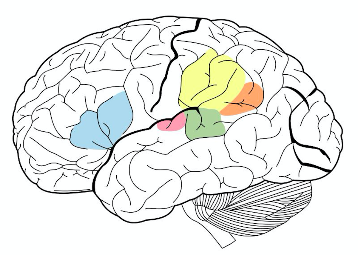


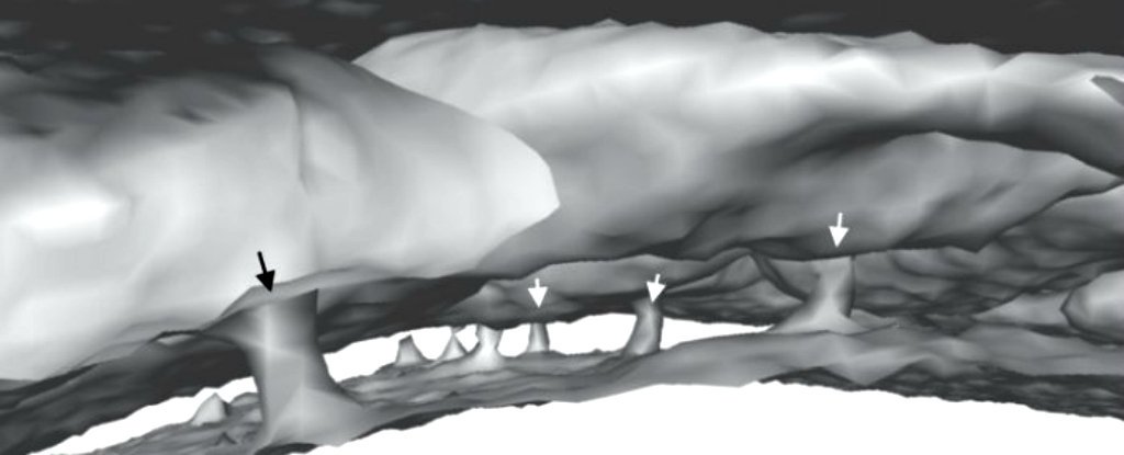
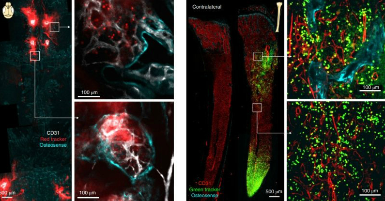 (Herrison et al./Nature Neuroscience)


 (Sirjhun Patel/BMJ Case Reports)


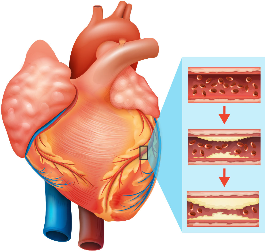


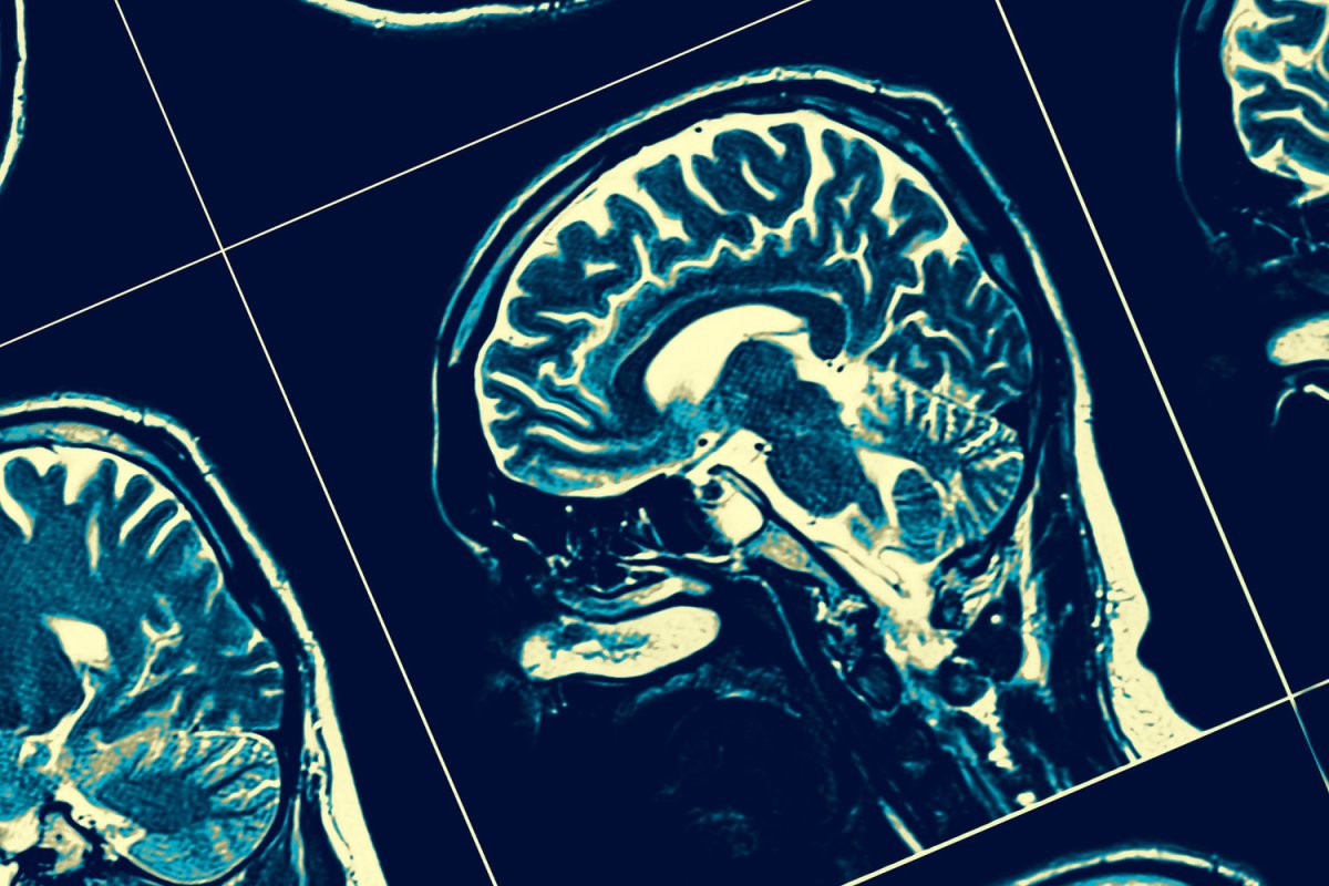


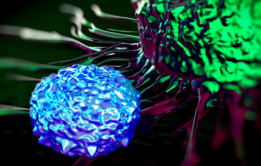


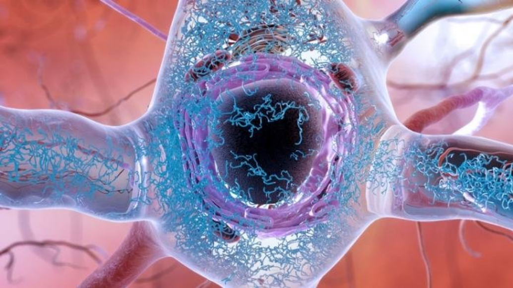

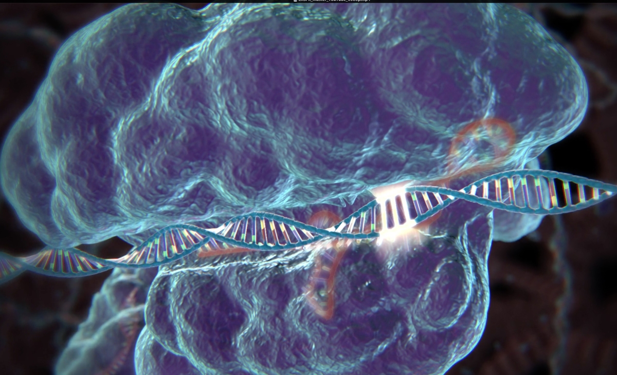


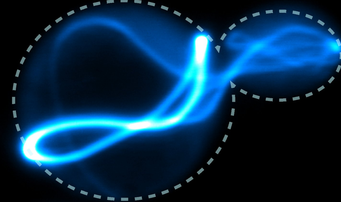

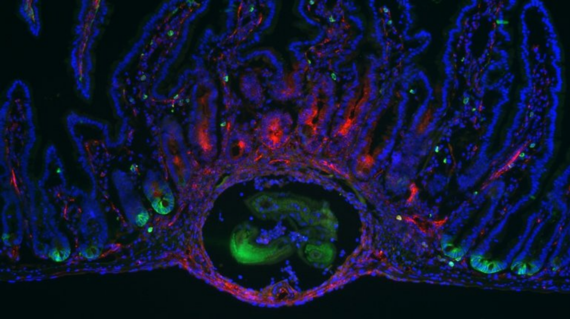

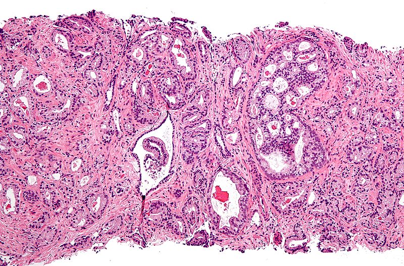
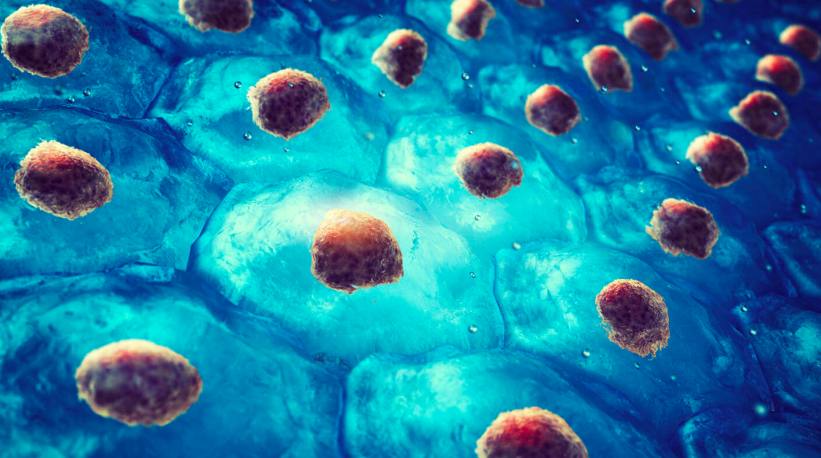


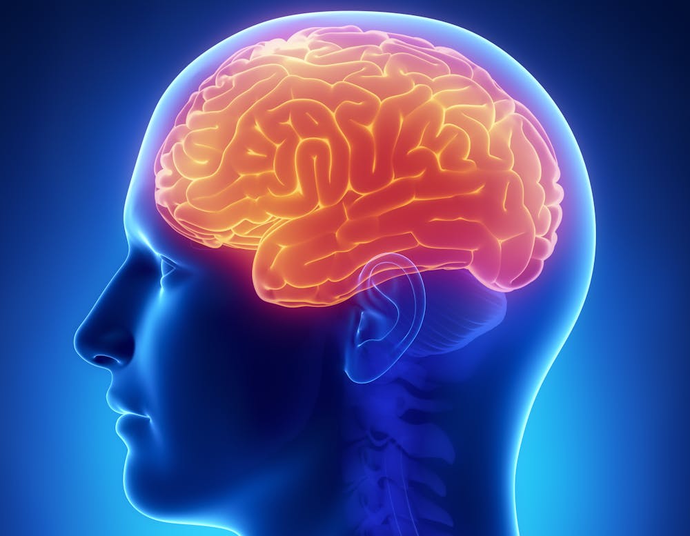
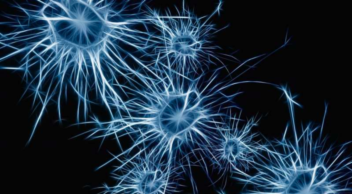


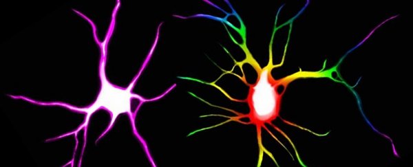
 The effects of drugs and one control (VEH) on neurons. (Ly et al., CC BY-ND)


 To see any protective effect in the brain in a clinical trial is completely new, and it supports the new theory that Alzheimer’s and Parkinson’s disease are caused, at least in part, by a lack of growth factor activity in the brain. These new theories bring a fresh view on how these diseases develop and increase the likelihood of developing a drug treatment that makes a difference.

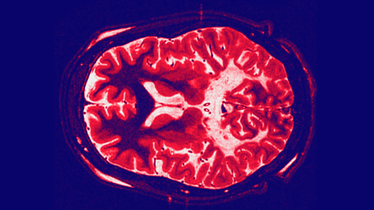
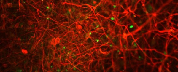
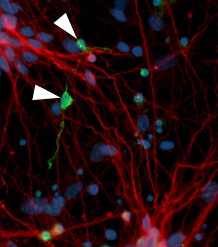 Neurofibrillary tangle-like structures (green) in affected neurons (red) (D Ovchinnikov)

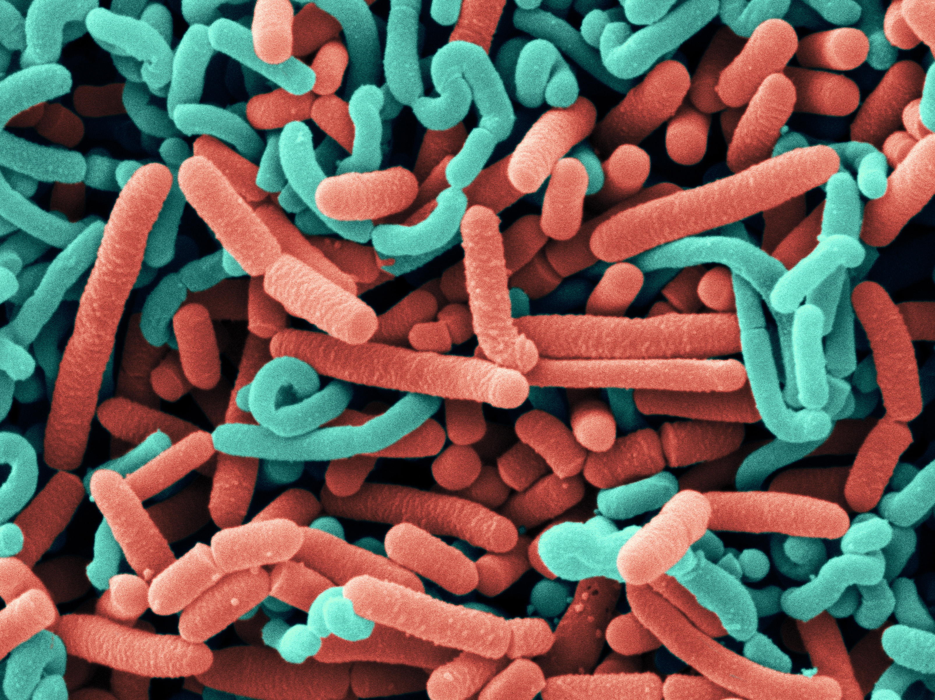